• Skip to primary navigation
  • Skip to main content
  • Skip to primary sidebar

Hip and Knee News

News Resource About Hip, Knee and Orthopedic Surgery

  • Home
  • Hip Surgery
  • Knee Surgery
  • Resources
    • Hip and Knee Glossary
  • About/Contact

Blog

OrthoTrophix Presents New Data Suggesting Joint Bone Shape as a Possible Surrogate Marker for Virtual Joint Replacement in Knee Osteoarthritis

by

FOSTER CITY, Calif., April 8, 2022 /PRNewswire/ — OrthoTrophix, Inc., a privately held biopharmaceutical company, announced today that the Company and its collaborators present clinical data strongly supporting joint bone shape change as part of a surrogate marker predictive of joint replacement in knee osteoarthritis (OA) patients.

An abstract entitled, “Improved WOMAC Physical Function is Associated with Slowed Pathological Bone Shape Change after TPX-100: Towards a Surrogate Marker for Virtual Knee Replacement?” was presented today in a Plenary Session of 2022 OARSI World Congress on Osteoarthritis in Berlin, Germany (Abstract 26 in Osteoarthritis and Cartilage Vol. 30 Suppl. S28–S29).

Functional impairment is a key risk factor for knee replacement even after adjusting for knee pain severity, based on the large NIH-sponsored Multicenter Osteoarthritis Study (the “MOST” study) involving over 5,500 knees. In the TPX-100-5, a Phase 2 study, placebo-treated knees with more advanced pathological joint bone shape change at baseline showed much faster progression of pathological bone shape change and poorer knee function at the end of the 12-month study period. In marked contrast, TPX-100-treated knees demonstrated reduction in pathological bone shape change and robust improvement of knee function through 12 months, regardless of severity at baseline. Consequently, both clinical (knee function) and structural (bone shape) efficacies of TPX-100 as compared to placebo were confirmed including in subjects with moderate to severe knee OA.

“The current FDA draft guidance indicates that a positive effect on an imaging marker of OA must be associated with avoidance or delay of the need for joint replacement, or must persuasively reduce deterioration of function and worsening of pain. This is a high bar,” commented Dr. Dawn McGuire, OrthoTrophix’ Chief Medical Officer. “However, the Multicenter Osteoarthritis Study findings and our clinical data collectively suggest that concordant improvements of bone shape change and clinical function could lead to a delay or elimination of the need for joint replacement surgery. These combined outcomes in structure and function could provide a ‘virtual joint replacement’ measure for the study of disease-modifying agents in individuals afflicted with knee OA.”

The Company also will present an abstract entitled, “Intra Articular TPX-100 Significantly Improves Pain Measures and Slows Pathological Bone Shape Chage in Knee OA”. This presentation demonstrates clinically meaningful improvements in overall knee pain and in specific key pain parameters in moderate to severe knee OA, linked to significant reductions in pathological bone shape change (Abstract 254 in Osteoarthritis and Cartilage Vol. 30 S193).

About OrthoTrophix, Inc.
OrthoTrophix, Inc., based in the San Francisco Bay Area, California, is a privately held biopharmaceutical company focused on development and commercialization of a first-in-class Disease Modifying Osteoarthritis Drug (DMOAD). Founded by three co-founders in 2011, the primary focus of OrthoTrophix has been regeneration and repair of cartilage and underlying bones in the knee and other joints with its novel proprietary compounds.

This press release contains “forward-looking” statements. These statements involve risks and uncertainties, which may cause results to differ materially from those set forth in the statements. The forward-looking statements include statements regarding product development and cannot be guaranteed. OrthoTrophix undertakes no obligation to publicly update any forward-looking statement, whether as a result of new information, future events, or otherwise. Forward-looking statements in this press release should be evaluated together with the many uncertainties that affect OrthoTrophix’ business.

Company Contact
Yoshi Kumagai
President and CEO
Tel: (510) 488-3824

SOURCE OrthoTrophix, Inc.

###[ad_2]
Originally Appeared Here
Filed Under: joint replacement, ORTHO NEWS

Over Two-Thirds of Hospitals are Noncompliant with Joint Replacement Price Transparency

by

[ad_1]

Despite CMS implementing a price transparency mandate, most hospitals are failing to comply when it comes to Total Joint Arthroplasty (TJA) procedures.

Hospitals are falling short of compliance on the price transparency mandate when it comes to two of the most common inpatient procedures.

According to a study by Clinical Orthopaedics and Related Research, only 32% of hospitals were fully compliant with price transparency on knee and hip replacement, or TJA.

When breaking down the results by individual procedures, 21% and 18% of hospitals provided the required information to be compliant for CPT codes 27447 and 27130, respectively, while 18% and 19% of hospitals did so for DRG codes 469 and 470.

Since the price transparency rule went into effect on January 1, 2021, CMS requires hospitals to provide five types of online price information for selected services: the total charge, the charges the hospital privately negotiated with health insurers, the minimum and maximum negotiated charges, and the amount the facility is willing to accept in cash.

Researchers of the study stated they chose to investigate price transparency for TJA procedures because of how widely used they are, making it some of the most commonly sought-after pricing information for orthopedic surgery patients.

The low compliance rates found in the study suggest hospitals are either struggling to implement the necessary changes to be compliant or are willfully ignoring the mandate.

“It is possible that hospitals have delayed compliance with the hope that related requirements may change or that current delays in enforcement may continue,” the authors wrote. “Although it is unclear whether legislation will change, the low rates of compliance demonstrated in our analysis should encourage hospitals to provide related information to avoid penalties expected to be enforced starting July 1, 2022.”

The study looked at 400 hospitals in December 2021 and searched each hospital’s website for a machine-readable file providing the five requirements for compliance. The researchers also considered hospitals pseudocompliant if they provided some type of gross price information.

The pseudocompliance rates were moderately higher, with 36% and 31% of hospitals offering total charges for CPT codes 27447 and 27130, respectively, and 34% and 50% of hospitals doing so for DRG codes 469 and 470.

Additionally, 13% of hospitals failed to provide machine-readable files, while 21% required users to provide personal information.

A recent JAMA study highlighted hospitals’ avoidance of price transparency, with the findings showing roughly 51% of facilities did provide either a machine-readable file or a shoppable display.

Only two hospitals, Northside Hospital Atlanta and Northside Hospital Cherokee, have so far been fined by CMS for failing to comply with price transparency requirements.

More fines are expected, but the authors of the TJA study believe hospitals should not only act to comply for the sake of their wallets, but also for the sake of their patients.

“Given the potential influence compliance and price sharing may have on empowering patients’ healthcare decisions and reducing healthcare expenditures in the United States, hospitals should use our analysis to identify where their compliance is lacking and to understand how to make their pricing information more readily available and comprehendible for the patients that they serve,” the researchers concluded.

Jay Asser is an associate editor for HealthLeaders.

###[ad_2]
Originally Appeared Here
Filed Under: ORTHO NEWS, ortho news - Google

Kinematic Alignment Bi-unicompartmental Knee Arthroplasty With Oxford Partial Knees: A Technical Note

by

Bi-unicompartmental knee arthroplasty (BiUKA) is an alternative to total knee arthroplasty for selected patients. Although it is thought to be technically demanding, the technique has not been previously described in detail. Kinematic alignment (KA) implantation and bone cuts parallel to the native joint line would be beneficial to ensure optimal mechanical loading. Here, we detail a technique for KA-BiUKA using the Oxford partial knees. The joint line is identified using the spoon of the microplasty instrumentation system with/without the accessory spoons. The tibia is cut parallel with the joint line using a side-slidable ankle yoke so that the inclination of the cutting block is parallel with the spoon surface. After defining the horizontal bone-cutting lines, the predominantly affected condyle is operated upon, followed by the lesser affected condyle. Although custom-made devices are required, the technique is useful and reproducible in the performance of KA-BiUKA with the Oxford partial knees.

Introduction

Unicompartmental knee arthroplasty (UKA) is an attractive surgery for unicompartmental knee osteoarthritis with functioning anterior cruciate ligament (ACL) [1,2]. It is characterized by quicker recovery, fewer systemic complications, lower postoperative mortality, and better range of motion than total knee arthroplasty (TKA) [3-5]. Another advantage of UKA is the retention of the ACL; once the ACL is sacrificed to facilitate a TKA, minor instability and alteration of kinematics are inevitable [6]. Unlike TKA, the original kinematics and joint stability can be retained in UKA, with improved patient satisfaction [7-9].

Despite these benefits, the usage of UKA depends on the integrity of the lateral compartment cartilage [10]. If the lateral compartment is damaged, conversion to TKA is unavoidable, even if the ACL is healthy. Bi-cruciate retaining (BCR) TKA is a possible alternative to conventional TKA, but it is a technically demanding procedure, and the results are not always consistent [6,11]. As described in the four-bar linkage theory, the ligament condition and morphology perfectly correspond to each other. If the morphology of the component matches the native morphology, the results can be excellent; otherwise, tightness and looseness inevitably emerge at certain angles. Complete replication of both medial and lateral components using existing TKA components is thus virtually impossible.

Bi-compartmental knee arthroplasty (BiUKA) is a potentially useful alternative to BCR because both compartments can be resurfaced individually [12-14]. Moreover, the kinematic alignment (KA) procedure is also possible if the components align with the original coronal joint line (CJL) obliquity. Performing KA-BiUKA with Oxford partial knees (OPKs) is also beneficial because the femoral components of OPK are partly spherical, meaning it can serve as a good imitation of the cylindrical axis. Moreover, bone cuts implemented along the CJL might be advantageous for the mechanical properties. Despite such benefits, however, there are no previous reports on BiUKA using OPKs (BiOUKA) except for its initial stage [15], where the surgical technique and instruments are immature and staged BiUKA for lateral compartment osteoarthritis after medial UKA [16]. We have modified the microplasty instruments to ensure tibial cuts parallel to the joint line. And there are no reports of KA-BiOUKA using additional components. This technical note describes KA-BiOUKA using custom-made instruments in detail.

Technical Report

Patient selection

The indication of KA-BiOUKA is functioning ACL and cartilage damage in both medial and lateral compartments. Full-thickness cartilage defects should be found in at least one compartment. In most cases, BiOUKA is a conversion from medial or lateral OPK owing to the intraoperative finding of cartilage damage on the opposite femoral condyle. BiOUKA is not applicable for severe patellofemoral joint diseases, such as bone defects and subluxation.

Preoperative radiographical planning

Preoperative anteroposterior radiography is used for planning. The medial and lateral joint lines are identified as the tangential line of the tibial articular surfaces. If both lines are straight and on the same level (leveled type; Figure 1, Panel a), the medial or lateral joint line is considered to be the CJL, and the single-spoon technique is used (described below). Otherwise, in the case of uneven type (Figure 1, Panel b), the double-spoon technique is performed with reference to the posterior condylar axis (PCA) intraoperatively. The predominantly affected condyle is determined, and this is operated first.

Figure
1:
Preoperative radiographic classification

(a) Leveled type: The medial and lateral tibial surfaces are aligned. In this type, the medial tibial surface represents the CJL obliquity. (b) Uneven type: The two tibial surfaces are not aligned. The posterior condylar axis is used to define the CJL obliquity.

CJL: Coronal joint line.

Joint opening

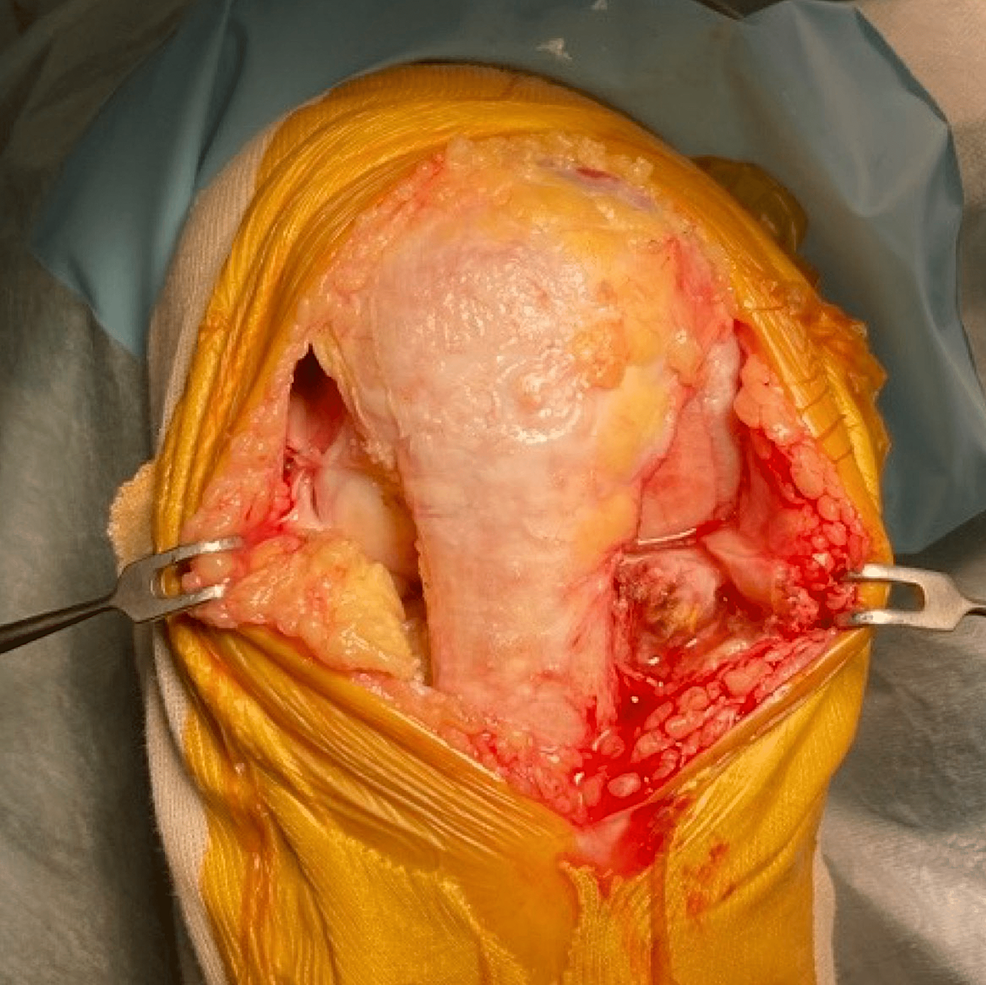
The medial parapatellar incision and medial parapatellar capsulotomy are performed for medial osteoarthritis (OA). Once the lateral cartilage lesion is found, the skin is peeled laterally so that the lateral border of the patella and patellar tendon is exposed. A lateral parapatellar capsulotomy is then added so that the lateral compartment can be manipulated (Figure 2). For the lateral OA, the lateral parapatellar approach is made and skin is peeled medially to expose the medial border of the patella and patella tendon, and this is followed by medial capsulotomy. Oxford mobile-bearing UKA is used for the medial side and fixed-lateral Oxford (FLO) is used for the lateral side. With the exception of the above-mentioned decision process regarding the tibial cutting plane, both procedures are implemented following the manufacturer-provided operation manuals [17].

Joint-opening

Figure
2:
Joint opening

For the medial OA, the medial parapatellar skin incision is used. The medial capsulotomy is implemented, and if lateral cartilage damage is found, the skin incision is extended proximally and slightly laterally. The skin flap is then peeled laterally to facilitate lateral capsulotomy.

OA: Osteoarthritis.

Deciding the tibial cutting plane

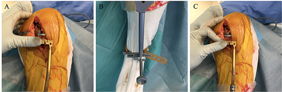
After joint opening and osteophyte removal, the tibial cutting plane is set parallel with the CJL. The single-spoon technique is used for leveled-type knees. The spoon gauge is inserted into the dominantly affected condyle, representing joint line inclination (Figure 3, Panel A). Our custom-made side-slidable ankle yoke is connected to the extramedullary (EM) rod instead of the original ankle yoke [18]. The sagittal inclination of the EM rod is adjusted so that it is parallel with the anterior cortex of the tibia. The cutting block is set just below the spoon. In most cases, the spoon is not parallel but rather varus to the cutting block (Figure 3, Panel B). The custom-made yoke is then slid laterally until the cutting block and the spoon are parallel (Figure 3, Panel C).

The-medial-spoon-technique-for-the-cutting-line-definition

Figure
3:
The medial spoon technique for the cutting line definition

(A) A spoon is inserted into the medial joint space. In straight-type knees, the inclination of the spoon represents the CJL inclination, the target of the cutting line. (B) The inclination of the tibial cutting block is different from that of the spoon. (C) The slide bar of the ankle yoke is adjusted so that the spoon and the cutting block are parallel.

CJL: Coronal joint line.

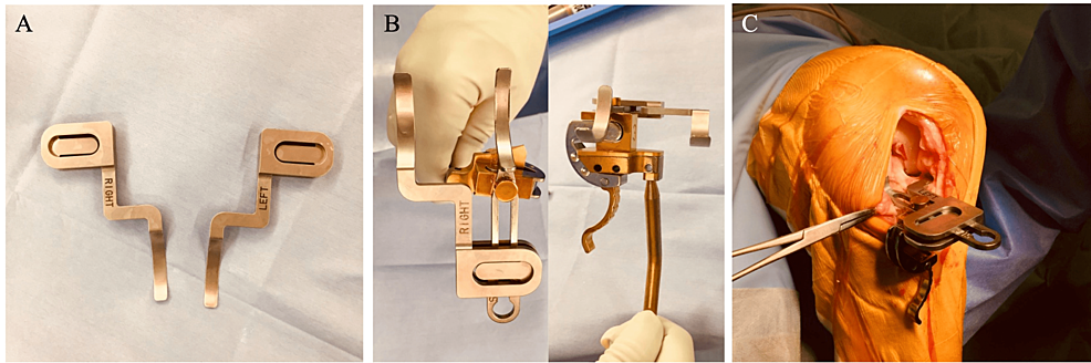
In non-straight-type knees, the double-spoon technique is performed with custom-made accessory spoons (Figure 4, Panel A). The accessory spoon is 0.5 mm thick and inserted into the opposite joint space, then incorporated with the conventional spoon. The spoons are thus set at the same level (Figure 4, Panel B). When both spoons are inserted into both compartments, it indicates the PCA (Figure 4, Panel C). The coronal alignment of the cutting block is adjusted as the single-spoon technique.

The-double-spoon-technique

Figure
4:
The double-spoon technique

(A) Accessory spoons. The accessory spoons are spoons of 0.5 mm thickness that can be joined with conventional spoons. (B) Once the accessory spoons are incorporated with the conventional spoon, both spoon levels are the same. (C) The inclination of the spoon indicates the posterior condylar axis when both spoons are inserted into the medial and lateral joint spaces.

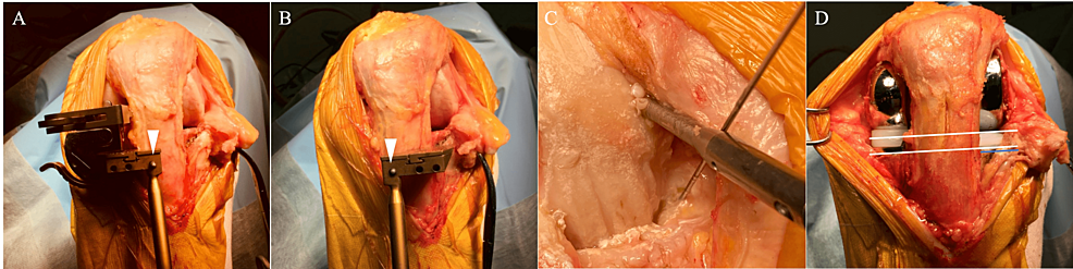
The spoon and the cutting block are fixed using the G-clamp, and the cutting block is fixed using a headless pin (Figure 5, Panel A). After the bone cut and adjustment of the flexion-extension gap are completed on the predominantly affected condyle, the cutting block is removed from the headless pin and changed to that on the opposite side using the same pin along with the extramedullary guide (Figure 5, Panel B) and the ankle yoke with retained extension, side-slide length, and posterior slope to maintain the cutting level as well as sagittal and coronal inclinations. The lesser affected compartment procedure is then performed. When the horizontal cuts are made, insertion of a K-wire at the tip of the tibial spine is recommended to prevent a horizontal overcut (Figure 5, Panel C). After the implantation, the CJL and cutting lines are virtually parallel (Figure 5, Panel D).

Bone-cutting-procedure-for-a-medial-dominant-osteoarthritis

Figure
5:
Bone-cutting procedure for a medial dominant osteoarthritis

(A) After the inclination and level of the cutting block are decided, a headless pin (arrowhead) is inserted into the tibia through the most lateral pinhole of the cutting block. The medial procedure is then performed. (B) The medial cutting block is changed to the lateral cutting block using the same pin connected to the yoke, and the same extent, slide, and posterior slope are maintained. (C) When performing the horizontal cut, a 2 mm K-wire is inserted to prevent a horizontal overcut. (D) The lateral procedure is then performed to set the medial and lateral cutting lines to be parallel.

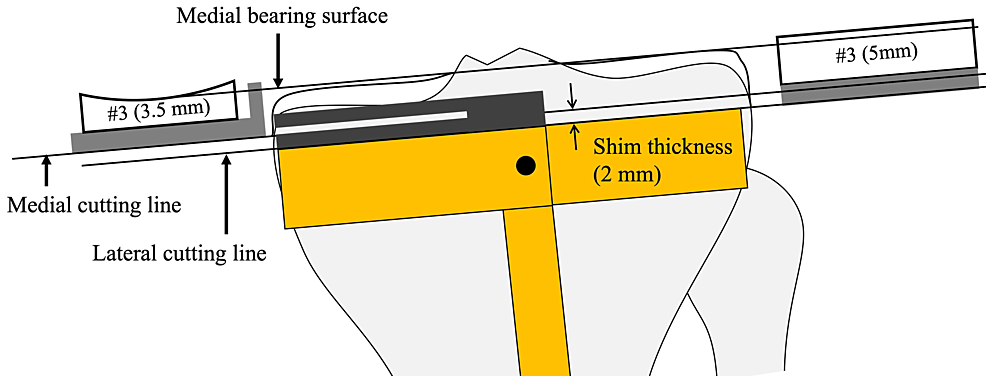
When the lateral tibial cuts are made, the shim is removed so that the cutting level is set to 2 mm lower than the medial cutting level. Both bearings are numbered, but the exact thicknesses of the bearings are 0.5 mm and 2.0 mm thicker than the labeled number. Complete leveling of both plateaus is therefore impossible when the level of the cutting block is constant (Figure 6). Eventually, the lateral CJL is inevitably 0.5 mm higher than the medial CJL, although this can be ignored.

Medial-and-lateral-tibial-cutting-level-in-the-kinematic-alignment-Oxford-unicompartmental-knee-arthroplasty

Figure
6:
Medial and lateral tibial cutting level in the kinematic alignment Oxford unicompartmental knee arthroplasty

The actual thicknesses are 0.5 mm and 2 mm thicker than the labeled number. Once the shim is removed, the lateral cutting level is 2.0 mm lower than that of the medial one. Eventually, the lateral bearing surface is 0.5 mm higher than the medial one when the same number bearing is used with the same cutting block level.

Postoperative radiographical evaluation

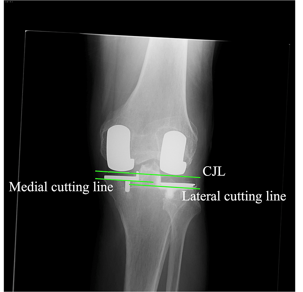
True anteroposterior radiography aligned to the tibial component surface is used for postoperative evaluation. Ideally, the postoperative CJL, which is the line tangential to both medial and lateral femoral components, is parallel with the medial and lateral cutting surface, and the medial cutting surface is 2 mm higher than the lateral cutting surface. In straight-type knees on the preoperative radiography, the CJL is expected to be parallel with the preoperative CJL (Figure 7).

Postoperative-radiography

Figure
7:
Postoperative radiography

The coronal joint line (CJL) is parallel to the cutting lines. Note that the lateral cutting line is lower than the medial cutting line.

Discussion

This is the first report to document KA-BiUKA by OPK in detail. Robotic-assisted BiUKA using a fixed-bearing component was recently reported, and constitutional whole leg alignment and joint line obliquity were shown to be restored [19]. Regarding OPK, it was used for BiUKA in the initial stage of the OPK [15]. Pandit et al. reported the staged BiUKA – adding a lateral UKA after medial UKA due to lateral compartmental osteoarthritis and showed satisfactory results [16]. More recently, a gait analysis showed that the subjects with BiUKA using OPK had similar gait characteristics to the normal subject compared to TKA subjects [20]. BiUKA has been reported to have mechanical advantages. A compression force on one component would cause a lift-off of the other component in the one-piece TKA component, but it never occurs in the two-piece tibial components in BiUKA [15,21]. The bone-cutting line was not shown in the previous studies; however, it is thought to play an important role in load transmission. A slight varus implantation of the tibial component was reported in previous biomechanical studies to reduce stress concentration in the medial tibial cortex, but a valgus placement increases it [22]. Although avoidance of valgus placement is important, the placement can be valgus against the proximal tibia in knees with tibia vara, which is especially prevalent in Asian patients [23,24]. Component placement parallel to the CJL might enable a proportioned load transmission across the joint.

In our technique, the dominantly affected compartment is operated upon prior to the lesser affected compartment. In this sequence, the operated condyle is always normal or nearly normal. By contrast, the procedure of the lesser affected condyle can be influenced by the disease of the opposite condyle, such as contracted or relaxed soft tissue and cartilage as well as bone loss. Our technique is a tibia-first sequence, in contrast with most KA-TKA techniques, in which the femur-first technique is used [25,26]. However, this is a standard technique in OPK and has been used for more than 40 years [27]. The tibia-first approach and incremental gap adjustment using the milling system can facilitate easy and precise adjustment of flexion and extension gaps. We believe the dominantly affected condyle-first and the tibial-first sequence might be ideal for facilitating the KA-BiOUKA.

There are some limitations in our report and technique. First, it was necessary to use custom-made devices (accessory spoons and side-slidable ankle yoke). Although a similar operation can be performed without the custom-made devices, where the cutting levels are decided individually using the standard spoon, the cutting plane is not parallel to the CJL. The CJL could be made parallel to the original CJL, but the kinematics and load distribution might be affected. Second, the lateral component is set in varus in the technique. This alignment is equivalent to a valgus placement of the medial UKA, which has reportedly increased the mechanical stress on the tibial cortex. Therefore, it can increase the risk of failure. Varus placement has not been reported to increase the risk of failure in lateral UKA. Third, there was no evaluation of clinical outcomes, in particular its superiority over TKA. A larger number of cases and long-term studies are needed to prove the benefits of the KA-BiUKA. Lastly, we used medial and lateral capsulotomy and disturbance of blood supply for the patella, followed by the avascular necrosis of the patella, and the anterior knee pain is a concern. The medial parapatellar approach for femoral and tibial bone cuts like TKA along with small lateral capsulotomy for lateral gap evaluation using the feeler gauge might be helpful. However, it might require additional instruments.

BiUKA is a great technically demanding operation; therefore, the establishment of the procedure is necessary for a fair evaluation of its effect. Our technique is considered to be easy and reproducible, so it can be implemented widely.

Conclusions

The details of an operative technique of BiUKA using OPK are presented as an alternative procedure for osteoarthritic knees with a functioning ACL and cartilage interaction on the lateral compartments. The technique can replicate the pre-arthritic joint line and maintain both cruciate ligaments, and a cylindrical axis is completely constructed. Moreover, the bone-cutting surface can be set in parallel to the joint line, which might be beneficial to load transmutation.


###[ad_2]
Originally Appeared Here
Filed Under: joint replacement, ORTHO NEWS

Will Exactech Joint Replacement Lawsuits Be Consolidated?

by

[ad_1]

On June 14, 2022, a group of plaintiffs filed a motion with the U.S. Judicial Panel on Multidistrict Litigation (JPML) to consolidate all Exactech joint replacement lawsuits in the U.S. District Court for the Eastern District of New York.

The plaintiffs claim that each of the actions involves common questions of fact and that consolidation would “serve the convenience of the parties and witnesses,” and “promote the just and efficient conduct of the litigation.”

Exactech Recalls Knee and Ankle Replacements Because of Insert Problems

In February 2022, Exactech issued a recall that impacted more than 140,000 Opetetrak, Optetrak Logic, and Truliant knee replacement systems as well as an additional 1,500 Vantage ankle replacements.

All of these devices used polyethylene inserts that according to Exactech, were packaged in non-conforming vacuum bags that lacked an additional protective layer. These bags could allow air to come into contact with the plastic inserts, which could cause oxidation and increase the risk that the devices would fail once implanted into a patient’s body.

Exactech has had similar problems with some of its hip implants, including the Connexion, Novation, and Acumatch. These have also shown a higher risk of premature failure because of problems with the polyethylene liner.

Plaintiffs Claim Faulty Inserts Led to Premature Implant Failures

According to the motion to transfer, there are already 27 Exactech lawsuits pending in 11 district courts across the country, with seven of those pending in the Eastern District of New York.

All of the cases involve similar allegations, with plaintiffs claiming that they received an Exactech knee or hip implant including polyethylene components that failed prematurely. The motion seeks to consolidate all of these cases—and any involving Exactech ankle replacements—into one court for pre-trial proceedings.

The Exactech recall includes polyethylene inserts manufactured as early as 2004 and packaged in out-of-specification vacuum bags that didn’t contain a second barrier layer. When these inserts fail, plaintiffs have to undergo revision surgery to remove the failed insert as well as other components, depending on the extent of the damage caused by the premature wear.

Degradation of the polyethylene can result in component loosening, tissue damage, osteolysis, permanent bone loss, and other injuries leading to complex revision surgeries and extensive recovery times.

Knee, Hip, and Ankle Devices All Share Similar Problems

The devices at issue for this proposed consolidated litigation include:

  • Opetrak Tibial Insert (knee implant)
  • Truliant Tibial Insert (knee implant)
  • Vantage Tibial Insert (ankle implant)
  • Connexion GXL Acetabular Liner (hip implant)

All of the liners that are included with these implants have been recalled. The plaintiffs seeking consolidation note that the cases involving these different implants are all related because their failures can be traced to the faulty polyethylene liner.


###[ad_2]
Originally Appeared Here
Filed Under: ORTHO NEWS, ortho news - Google

Video Evidence of Tissue Sliding Improvement by Ultrasound-Guided Hydrorelease on Scars After Arthroscopic Knee Surgery: A Case Report

by

Postoperative scarring is a complication of arthroscopic knee surgery that causes a lack of terminal extension and tissue sliding defects. We present video evidence of tissue sliding before and after ultrasound-guided hydrorelease in a 53-year-old man. The patient presented with pain in the scarred area following arthroscopic knee surgery. His active and passive extension was -5° with restricted patellar mobility. Dynamic ultrasonography revealed scar tissue sliding defects. For ultrasound-guided hydrorelease, a needle (22G, 60 mm) was aimed at a site within 10 mm depth of the hypoechoic change in the scar area below the patella, and saline solution (10 mL) mixed with 1% lidocaine (10 mL) and 10 mg prednisolone was injected. Immediately after injection, the patient’s extension was 0° with no pain or limitation of patellar mobility, and dynamic ultrasonography showed tissue sliding improved. Video evidence from dynamic ultrasonography clarifies the direction of the inadequate slide and the indication for and efficacy of ultrasound-guided hydrorelease. This case highlights the benefits of video evidence from dynamic ultrasonography before and after ultrasound-guided hydrorelease.

Introduction

Postoperative scarring is a complication of arthroscopic knee surgery and contributes to progressive fibrosis in the infrapatellar fat pad, lack of terminal extension, anterior knee pain (AKP), and decreased patellar mobility [1,2]. The proposed treatments for this issue are anterior interval release [3,4] and manual therapy [5], although multiple arthroscopic surgeries may lead to additional complications. A less invasive treatment than arthroscopic surgery would be beneficial for patients suffering from these types of pathologies.

Ultrasound-guided hydrorelease has been performed for lower back pain [6] and postoperative scar tissue [7]. Machida et al. consider ultrasound confirmation of improved tissue sliding as the key to the success of this procedure regarding the treatment of scar tissue after arthroscopic surgery. However, no video evidence of improved tissue sliding has been presented [7]. We performed an ultrasound-guided hydrorelease in a patient who presented with a lack of terminal extension and AKP after arthroscopic surgery for medial meniscus injury. We present video evidence of tissue sliding before and after the procedure in this patient.

Case Presentation

A 53-year-old man (height: 170 cm; body weight: 80 kg; body mass index: 27.7 kg/m2), diagnosed with medial meniscus injury, presented with AKP after previous arthroscopic knee surgery. He developed left knee pain six months before surgery and underwent arthroscopic medial meniscus repair after conservative therapy failed to improve his pain. Arthroscopic surgery was performed according to the standard all-inside repair technique using FasT-Fix system (Smith & Nephew Endoscopy, Andover, USA) [8] (Figure 1), and range-of-motion exercise was started after two weeks of knee brace immobilization. However, his range of motion remained limited even at six weeks postoperatively, and the scar under the patella tightened during extension, causing him severe pain. Active and passive extension was -5° with restricted patellar mobility, passive flexion was 115°, and an 11-point numerical rating pain scale (NRPS; 0 (“no pain”) to 10 (“worst possible pain”)) was three in walking. The Knee injury and Osteoarthritis Outcome Score (KOOS) showed KOOS pain of 25.0, KOOS symptoms of 17.9, and KOOS activities of daily living (ADL) of 47.0 (Table 1) [9].

Figure
1:
Arthroscopic views in the patient’s left knee

A) Before repair: Horizontal tear of the medial meniscus (arrow). B) After repair: Horizontal tear of the medial meniscus repaired by the all-inside repair technique (arrow).

  Six weeks postoperative, before hydrorelease Seven weeks postoperative, after hydrorelease 15 weeks postoperative
Knee extension, degree -5 0 0
Knee flexion, degree 115 120 145
Pain in walking, NRPS 3 1 1
KOOS pain 25 N/A 66.7
KOOS symptoms 17.9 N/A 57.1
KOOS ADL 47 N/A 91.0
Table
1: The patient’s range of motion and function course

NRPS: Numerical rating pain scale; KOOS: The Knee injury and Osteoarthritis Outcome Score; ADL: Activities of daily living; N/A: Not available.

The dynamic ultrasonography six weeks postoperatively revealed scar sliding defects during quadriceps contraction (Video 1) and manual skin sliding (Video 2). We then performed ultrasound-guided hydrorelease, as reported by Machida et al. [7].

Video
1:
Dynamic ultrasonography at medial scar area during quadriceps contraction

A) Before ultrasound-guided hydrorelease: The hypoechoic area is present in the scar area. Tension from quadriceps contraction is not transmitted distally (arrow). B) After ultrasound-guided hydrorelease: Tension from quadriceps contraction is transmitted distally beyond the hypoechoic area of the scar (arrow).

Video
2:
Dynamic ultrasonography at medial scar area during manual skin surface sliding

A) Before ultrasound-guided hydrorelease: Manually sliding over the skin surface does not slide over the hypoechoic areas of the scar (arrow). B) After ultrasound-guided hydrorelease: The movement of the scar area during manual sliding on the skin surface is greater than before ultrasound-guided hydrorelease (arrow).

For the ultrasound-guided hydrorelease, the patient was positioned in 30° flexion of the knee joint in the supine position. Ultrasonography was performed using a three to 11 MHz B-mode linear array scanner (SONIMAGE MX1, Konica Minolta, Tokyo, Japan). For ultrasound-guided hydrorelease, a needle (22G, 60 mm) was aimed at a site within 10 mm depth of the hypoechoic change in the scar area below the patella, and saline solution (10 mL) mixed with 1% lidocaine (10 mL) and 10 mg prednisolone was injected: first on the lateral scar below the patella and then the same for the medial scar the following week, and Video 1 and Video 2 show the medial scar. Only one injection was performed for each, and the treatment time was approximately 10 minutes, including an ultrasound examination. All procedures were performed by one orthopedic surgeon with more than 10 years of experience.

Immediately after second injection, left knee extension was 0° without pain, flexion was 120°, and pain in walking was a score of one on the NRPS. Dynamic ultrasonography showed that scar tissue sliding improved (Video 1 and 2). After that, rehabilitation continued with a range of motion exercise, manual therapy [5], mobilization of the patella, and muscle strengthening. At 15 weeks postoperatively (i.e., at nine weeks post hydrorelease), his left knee extension was 0° with no pain or limitation of patellar mobility, flexion was 145°, and pain in walking was one on NRPS. KOOS pain, symptoms, and ADL scores were 66.7, 57.1, and 91.0, respectively. After the injections, the patient felt heaviness in the left knee for one day, which improved spontaneously without any specific treatment. After that, the heaviness never recurred. He was satisfied with the ultrasound-guided hydrorelease and subsequent rehabilitation and could return to work as an auto mechanic.

Discussion

The most important finding from this patient’s course was that ultrasound-guided hydrorelease to the scar after previous arthroscopic knee surgery improved the terminal extension with AKP. Furthermore, the video evidence showed that scar tissue sliding had indeed improved.

The patient’s terminal extension was ultimately improved by ultrasound-guided hydrorelease. A previous case series on arthroscopic anterior interval release showed that all patients with a lack of terminal extension had improved [10]. We believe ultrasound-guided hydrorelease may also be effective, as this patient’s progress demonstrates. The advantage of this procedure is that it is less invasive than arthroscopic anterior interval release, which may be beneficial for postoperative patients.

Notably, the video evidence objectively confirmed that scar tissue sliding improved. This highlights the effectiveness of dynamic ultrasonography. The video evidence also identifies the stimulus and direction of the sliding defects. With video evidence, we can clearly visualize the indications and results of ultrasound-guided hydrorelease. Evaluation of the anterior interval scar is recommended for patients suffering from lack of terminal knee extension, AKP, and decreased patellar mobility, which may be indications for arthroscopic release [4]. We recommend that surgeons confirm the scars in patients presenting with similar symptoms and attempt dynamic ultrasonography and ultrasound-guided hydrorelease before arthroscopic anterior interval release.

In a previous report by Machida et al. [7], who proposed this technique, the mechanism of tissue separation by ultrasound-guided hydrorelease may have improved tissue sliding. Therefore, confirmation of the sliding by ultrasonography will be the key to the success of this procedure. To the best of our knowledge, this is the first report to present video evidence of improved tissue sliding before and after ultrasound-guided hydrorelease for arthroscopic scars.

Ultrasound-guided hydrorelease has several strengths. It can immediately improve terminal knee extension in patients with AKP. Also, because of its less invasive procedure, knee brace immobilization or unweighting are not necessary, and exercise therapy can begin immediately. Another major strength is that the procedure is safe. The patient had no serious adverse events requiring specific treatment. Furthermore, no serious adverse events have been reported in previous studies [6,7,11]. Finally, ultrasound-guided hydrorelease can be presented on videos, which can aid in informing the patient before obtaining the patient’s consent for the procedure.

Despite these benefits, there are still some unknowns with ultrasound-guided hydrorelease. First, the timing of its application is unknown. In a previous study by Machida et al. [7], the patient was in the tenth month after arthroscopic knee surgery, whereas in this case report, the patient was in the sixth week after surgery. The second is an injection agent. We injected saline mixed with lidocaine and prednisolone because the patient felt severe pain. In previous studies, saline only [6,7], saline mixed with 0.5% mepivacaine hydrochloride [11], and bicarbonate Ringer’s solution [11] have been used. In a comparison of the analgesic effects of injectable agents, saline alone has been reported to be more analgesic than local anesthesia. [12].

Conclusions

Ultrasound-guided hydrorelease applied to the scar after arthroscopic surgery resulted in full extension of this patient’s knee joint without pain and video evidence of improved sliding was presented. Video evidence from dynamic ultrasonography has the advantage of clarifying the direction of the lack of sliding and the indication for and efficacy of ultrasound-guided hydrorelease. This case highlights the benefits of video evidence from dynamic ultrasonography before and after ultrasound-guided hydrorelease.


###[ad_2]
Originally Appeared Here

Filed Under: KNEE, Knee Surgery, ORTHO NEWS

American Joint Replacement Registry Releases 2021 Annual Report, Showing Increase in Number of Hip and Knee Procedures Despite Pause Due to COVID-19

by

ROSEMONT, Ill., Nov. 12, 2021 /PRNewswire/ –The American Joint Replacement Registry (AJRR), the cornerstone of the American Academy of Orthopaedic Surgeons (AAOS) Registry Program, released its 2021 Annual Report on hip and knee arthroplasty procedural trends, and patient outcomes today at the American Association of Hip and Knee Surgeons’ (AAHKS) 2021 Annual Meeting. Despite the disruption to the delivery of joint replacement care during the initial impact of the COVID-19 pandemic (March through May 2020), procedures rebounded to historic averages by June 2020. Even with the temporary decline in procedures, the report demonstrates an overall cumulative procedural volume growth of 18.3% compared to the previous year and includes findings from 2,244,587 hip and knee arthroplasty procedures performed between 2012 and 2020.

The American Joint Replacement Registry 2021 Annual Report shows an increase in the number of hip and knee procedures despite pause due to COVID-19 pandemic.

“The ability to return to normal procedural volume rates just a few months after the COVID-19 pandemic began is a testament to the commitment and resiliency of healthcare institutions, clinicians and patients,” said Bryan D. Springer, MD, FAAOS, chair of the AJRR Steering Committee. “While we are experiencing unprecedented times in healthcare and continue to navigate the challenges of the pandemic, the AJRR remains committed to the growth and expansion of the registry to paint a more complete picture of our patient population. Through increased participation and a successful integration of Medicare claims data, we are providing orthopaedic surgeons, hospitals and stakeholders with actionable data, insights and trends to improve the lives of millions of Americans who suffer from hip and knee arthritis.”

AJRR is the largest orthopaedic registry in the world based on annual procedures submitted, and the 2021 AJRR Annual Report marks the eighth annual report. With the collection and reporting of U.S. hip and knee arthroplasty data, the report aims to provide valuable information to orthopaedic surgeons, hospitals, ambulatory surgery centers, private practices, device manufacturers, payers, and most importantly patients. The 2021 report represents over 2.2 million hip and knee procedures from over 1,150 hospitals, ambulatory surgery centers (ASCs), and private practice groups submitting data from across all 50 states and the District of Columbia.

James A. Browne, MD, FAAOS, chair of the AJRR Publications Subcommittee and editor of AJRR Publications added: “This year’s AJRR Annual Report provides the most comprehensive picture to date of patterns of hip and knee arthroplasty practice and outcomes in the United States. For the first time this year, cumulative percent revision curves were produced with a diagnosis-specific endpoint examining revision due to infection for total knee arthroplasty (TKA) and revision due to periprosthetic fracture for total hip arthroplasty (THA) patients over 65 years of age. The registry continues to use more sophisticated and detailed survivorship curves, including device-specific cumulative revision stratified by bearing and fixation type, in addition to utilizing Centers for Medicare & Medicaid Services (CMS) data.”

Additional findings from the 2021 AJRR Annual Report include:

  • Device-specific revision analyses showed that all included hip device constructs had a cumulative percent revision of less than 2.8% at one year and less than 4.7% at final follow up for each respective device. All knee device constructs included in analysis had a cumulative percent revision of less than 2.5% at three years and less than 3.7% at final follow up for each respective device.  
  • The use of cement for femoral component fixation is slowly increasing for both elective primary THA as well as arthroplasty for femoral neck fracture, and cementless fixation shows a statistically significant reduction in early revision due to periprosthetic fracture, compared to cementless fixation in elective primary THA patients over 65 years of age.
  • While cemented fixation for TKA still predominates, the report shows that cementless fixation continues to increase and was associated with significantly less revision due to infection in elective primary TKA patients over 65 years of age.
  • For both TKA and THA procedures, postoperative length of stay continues to decrease.

Enhanced Registry capabilities offerings and include:

  • Additional opportunities for sites to track performance measurements and use Registry data in national quality improvement (QI) programs.  
  • A 39% increase in sites reporting patient-reported outcome measures (PROMs), compared to the previous year, through continued support of the RegistryInsights® PROM platform and partnerships with third-party vendors with the expanded Authorized Vendor Program.
  • Peer-reviewed publications and presentations based on AJRR Registry data.

To read and download the complete 2021 report, visit the AJRR website. Slides with figures and data tables as featured in the report are also available.

AAOS Registry Program 
The AAOS Registry Program’s mission is to improve orthopaedic care through the collection, analysis, and reporting of actionable data. The American Joint Replacement Registry (AJRR), the Academy’s hip and knee replacement registry, is the cornerstone of the AAOS’s Registry Program, and the world’s largest national registry of hip and knee joint replacement data by annual procedural count, with more than 2.4 million procedures contained within its database. Additional registries include the Fracture & Trauma Registry, the Musculoskeletal Tumor Registry (MsTR), the Shoulder & Elbow Registry (SER), and the American Spine Registry (ASR), a collaborative effort between the American Association of Neurological Surgeons (AANS) and the AAOS. 

About the AAOS
With more than 39,000 members, the American Academy of Orthopaedic Surgeons is the world’s largest medical association of musculoskeletal specialists. The AAOS is the trusted leader in advancing musculoskeletal health. It provides the highest quality, most comprehensive education to help orthopaedic surgeons and allied health professionals at every career level best treat patients in their daily practices. The AAOS is the source for information on bone and joint conditions, treatments, and related musculoskeletal health care issues, and it leads the health care discussion on advancing quality.

Follow the AAOS on Facebook, Twitter, LinkedIn, and Instagram.

SOURCE American Academy of Orthopaedic Surgeons

Related Links

www.aaos.org


###[ad_2]
Originally Appeared Here

Filed Under: joint replacement, ORTHO NEWS

Lose Weight, Slow Knee OA Progression? New Study Suggests Yes

by

Declines in body mass index (BMI) were linked with slower worsening of knee osteoarthritis (OA), according to data from three large longitudinal cohort studies.

With more than 6,000 knees evaluated for OA progression and 4 to 5 years of follow-up, “each 1-unit decrease in BMI was associated with a 4.76% reduction … in the odds of the incidence and progression of the overall structural defects of knee osteoarthritis” as assessed under the Kellgren-Lawrence grading system, reported Zubeyir Salis, BEng, of the University of New South Wales in Kensington, Australia, and colleagues in Arthritis & Rheumatology.

While such a relationship seems obvious, it has evaded firm proof, the researchers noted. Earlier studies had tied body weight to risk of OA development and the likelihood of progression and end-stage outcomes such as total joint replacement. But whether losing or gaining weight alters the trajectory over time is another matter.

Salis and colleagues identified just one previous attempt to address the question directly: patients already showing joint degeneration who lost around 10 kg (22 lb) on average in a randomized diet-and-exercise trial showed no less progression than a control group with little weight loss, but follow-up only lasted 18 months. (A number of other studies had lumped patients without structural damage at baseline together with those with established degeneration, and they had mixed results.)

For the current analyses, Salis and colleagues combined data from three independent cohort studies from the U.S. and the Netherlands: the Osteoarthritis Initiative (OAI), which was also the basis for some of the above-mentioned studies; the Multicenter Osteoarthritis Study (MOST); and the Cohort Hip and Cohort Knee (CHECK) study. These data covered 9,683 knees (5,774 individual patients) for assessing the incidence of structural knee degeneration (i.e., no evidence of joint damage at baseline) and 6,074 knees (3,988 individuals) for progression of established degeneration.

Structural joint parameters were measured with x-rays at baseline and at follow-up, which was 4 years in the OAI and 5 years in the other two cohorts. Changes in these parameters (medial and lateral joint space narrowing and femoral and tibial surface osteophytes) were correlated against changes in BMI.

For patients included in the evaluation of new-onset structural damage, mean age at baseline was 60; about 40% were men and 88% were white. BMI at baseline averaged 28.2, with 33% of participants classified as obese (BMI ≥30). A total of 1,101 participants in this analysis saw BMI declines of at least 1 unit during follow-up, whereas 1,611 had increases of 1 or more units.

Patients’ baseline characteristics in the study of progression were similar. Baseline BMI was a bit higher on average (30.4), and 48% were obese. BMI declines of 1 or more units were seen in 798 participants, while 1,008 had increases.

Salis and colleagues found a significant association between BMI changes and the risk of developing structural damage, with an odds ratio of 1.05 (95% CI 1.02-1.09) for each 1-unit increment in BMI. This value was nearly identical to that seen for risk of progression in patients with damage at baseline (OR 1.05, 95% CI 1.01-1.09). In both analyses, joint space narrowing in the medial area largely drove the overall findings, with odds ratios in both cases of 1.08 per 1-unit BMI increment.

However, the results suggested that weight loss was far from the only factor at play in governing joint-damage incidence and progression. The researchers estimated population-attributable fractions of the total risk accounted for by weight loss at 13% for incidence and 10% for progression.

In addition, the investigators stopped short of asserting that the findings prove that weight loss slows OA disease progression, saying they “showed evidence of association, not causality.” But the researchers did argue that “people with overweight or obesity — and potentially also those of normal weight — may benefit from a decrease in BMI to prevent, delay or slow the structural defects in knee osteoarthritis.”

  • John Gever was Managing Editor from 2014 to 2021; he is now a regular contributor.

Disclosures

This study was supported by the Australian government; the individual cohort studies each had their own funding sources, none of which were commercial entities.

Salis and one co-author were co-owners of a company dedicated to education around weight management. Two co-authors also reported relationships with multiple pharmaceutical companies.

###[ad_2]
Originally Appeared Here

Filed Under: joint replacement, ORTHO NEWS

Five ways to tackle the joint pain of arthritis

by

[ad_1]

So what are the other options?

1. Keep moving

It’s probably the opposite of what you want to do, and of what your osteoarthritis seems to be telling you, but the evidence is rock-solid. Exercise helps in multiple ways. “It improves nutrition and blood flow to the joint, lines up the joints, strengthens muscles, improves stability and restores function,” says Dr Benjamin Ellis, consultant rheumatologist and senior clinical advisor for Versus Arthritis. Avoiding activity because of osteoarthritis pain kicks off a vicious “deconditioning cycle”. Muscles weaken, joints become less stable and the pain gets worse. Exercise also helps with weight control, which is critical. “For every pound you carry, four times that amount will go to your weight bearing joints,” says Dr Wendy Holden, honorary consultant rheumatologist at North Hampshire Hospitals NHS Foundation Trust and medical advisor of Arthritis Action. “If you’re just 10kg overweight, that’s another 40kg to bear.” 

Experts advise that any exercise is good, even gardening. “Whatever you enjoy, whatever you’re doing, do more,” says Holden. “A recent study found that just walking will reduce the pain from early knee osteoarthritis and also make it much less likely to progress – so it’s potentially preventative.”

Versus Arthritis has an online programme, Let’s Move With Leon, presented by fitness expert Leon Wormley and his mum Janet who lives with arthritis. There’s also Escape-Pain, a national group rehabilitation programme to reduce arthritic pain through exercise. 

2. Medical

The latest research suggests that paracetamol performs no better than a placebo for osteoarthritis, while strong and potentially addictive opioids bring more risk than benefits. The first best option, according to NICE, is topical NSAIDS (non-steroidal anti-inflammatory drugs). “Gels that you rub on your skin can work well and stay where you put them, on the joint, unlike oral NSAIDS which end up in the blood stream,” says Ellis. Capsaicin is also recommended by NICE – a cream which uses the compound from chilli peppers to numb pain over a period of time. “Capsaicin comes up very often on our helpline and has been the most frequent question in relation to alternative therapies,” says Zoë Chivers, director of services and influencing at Versus Arthritis. 

It’s not something you can use now and again. “You have to really go at it, applying four times a day for three or four weeks to get the effect,” says Ellis. 

Anti-inflammatories like ibuprofen can help, and weak opioids like codeine have a role for certain occasions – big trips, a challenging day ahead. Corticoid steroid injections bring relief for up to two or three months – the evidence is strongest for pain on hip joints.  

“If you have tried all these and can’t live with the pain, you’re heading into joint replacement territory,” says Ellis. Waiting lists there can be several years. The future – the holy grail – could be cartilage transplantation or the injection of stem cells to stimulate new cartilage growth. Research is ongoing, but both have been performed at the Royal National Orthopaedic Hospital in Stanmore.

3. Hot and cold 

###[ad_2]
Originally Appeared Here

Filed Under: ORTHO NEWS, ortho news - Google

Outpatient joint replacement vs. ambulatory joint replacement?

by

[ad_1]

Over the past 5 years, there has been a lot of discussion about “outpatient joint replacement.”

With all the emphasis on value-based care, joint replacement has been targeted as an
opportunity to reduce cost by movement from inpatient to outpatient admissions, resulting in a
reduction in skilled nursing admissions and decreased home health utilization. The BPCI
program led the way and continues to be successful in some markets. Many research articles
have been published in peer-reviewed journals citing the cost savings and the improvement in
outcomes. However, there still seems to be some hesitancy for migration to the ambulatory
surgery center. In 2016, the SG2 forecast was for 32% of joint replacements to be done in the
ASC by 2022. We haven’t even hit half of that. Why?

REASON #1: Site of Serve Shift. It is one thing to do an “outpatient joint” in the inpatient
setting where it is coded as HOPD (hospital outpatient department). You still have the safety net
of the big hospital; you still have your same staff, same rooms and it is, in fact, cheaper with a
reduced facility fee/DRG payment. However, to move it to the most cost-effective venue of the
outpatient surgery center it requires an entirely new care paradigm, from pre-admission testing
to patient education, staffing, sterile processing, care management, etc. So, the site of service
shift is not easy and, therefore, slow to progress and hit the SG2 forecast.

REASON#2: Alignment. To shift the site of service to the ASC, there must be alignment with
the surgeon, the payer and the ASC. Since it is a heavy lift to create programs and protocols
around a purely outpatient joint without an inpatient backdrop, systems have struggled to
figure it out and shift the volume to the ASC in large numbers. It is simply easier to keep doing
things the way you always have unless there is some incentive to change. Commercial
bundles and increased facility fees in physician-owned ASCs have seen excellent success in
certain markets.

REASON #3: Risk Assessment. The pandemic certainly gave outpatient joints a push and
many surgeons were forced to ask a different question as their OR’s were closed. We used to
ask “Who can I do at the ASC?” However, now we ask “Who CAN’T we do at the ASC?” To
help answer this question, we developed an evidence-based risk assessment tool, which is
built into our software program called ValereCARE, that we put all of our patients through. They
are assessed as type 1(healthy and safe to be done in the ASC), type 2 (comorbid conditions
needing clearances but can be done in the ASC if cleared) and type 3 (too many co-morbid
conditions and should be done in the hospital setting). We are embarking on a research study to
validate this tool and hope for others to be able to utilize it in their ASC’s.

We hope the industry progresses more and more towards ambulatory joint replacement as
surgeons and their staff get comfortable with this new value-based paradigm of care. It
requires alignment of payers, ASC executives and other industry leaders such as implant
companies. We will be gathering these various groups for the third time at our Valere Summit
on Ambulatory Joint Replacement on September 22-23 and all are welcome to come and take
part in this interactive value-based discussion. Registration and an agenda can be found at
www.valerebundledsolutions.com/summit.


###[ad_2]
Originally Appeared Here

Filed Under: ORTHO NEWS, ortho news - Google

Aspirin led to more dangerous blood clots in hip and knee replacements: study

by

[ad_1]

Taking aspirin after a joint replacement surgery could increase the risk of blood clots, according to a study published Aug. 23 in the Journal of the American Medical Association.

In the study, 9,711 patients who underwent hip and knee replacements were split into two groups. One group was treated strictly with aspirin after the surgery, while the other was only given the anticoagulant enoxaparin.

After 90 days, researchers evaluated both groups and found that venous thromboembolism occurred in 256 patients. The group treated strictly with aspirin were nearly twice as likely to develop VTE, according to the study.

The aspirin group saw a 3.45 percent VTE rate, while only 1.82 percent of the enoxaparin group experienced VTE.

###[ad_2]
Originally Appeared Here

Filed Under: ORTHO NEWS, ortho news - Google

  • « Go to Previous Page
  • Page 1
  • Page 2
  • Page 3
  • Page 4
  • Page 5
  • Page 6
  • Interim pages omitted …
  • Page 13
  • Go to Next Page »

Primary Sidebar

ORTHO NEWS

Aspirin led to more dangerous blood clots in hip and knee replacements: study

Spinal anesthesia tied to increased opioid use after hip surgery

Useful Health Tips for People With Chronic Hip Issues

Useful Health Tips for People With Chronic Hip Issues

A robotic assistant for joint replacement surgery

A robotic assistant for joint replacement surgery

Prior Diagnosis of COVID Has No Increased Complications in Total Joint Arthroplasty

Prior Diagnosis of COVID Has No Increased Complications in Total Joint Arthroplasty

Joint replacement: Myths and facts related to knee replacement surgery | Health

Joint replacement: Myths and facts related to knee replacement surgery

Learn More About Total Hip Arthroplasty and the Function of Robotics in THA

Learn More About Total Hip Arthroplasty and the Function of Robotics in THA

Am I Too Old to Get My Knee or Hip Replaced? What to Consider at Age 70, 80 and Up

Am I Too Old to Get My Knee or Hip Replaced? What to Consider at Age 70, 80 and Up

Putting Your Best Foot Forward for Joint Surgery Success | Health

Putting Your Best Foot Forward for Joint Surgery Success

Use of Antibiotic Lavage in Total Knee Replacement to Prevent Postoperative Infection

Use of Antibiotic Lavage in Total Knee Replacement to Prevent Postoperative Infection

Tips to care for an elderly after hip replacement surgery

Tips to care for an elderly person after hip replacement surgery

Copyright 2014 All Rights Reserved · DISCLAIMER: Nothing here constitutes legal, medical, or other advice; all content relates to an individual perspective only. A professional relationship with a physician, or with a lawyer is built over time, with mutual investment, trust, and respect. This site is not a substitute for that.
~ THIS DOMAIN IS FOR SALE ~

Privacy Policy