• Skip to primary navigation
  • Skip to main content
  • Skip to primary sidebar

Hip and Knee News

News Resource About Hip, Knee and Orthopedic Surgery

  • Home
  • Hip Surgery
  • Knee Surgery
  • Resources
    • Hip and Knee Glossary
  • About/Contact

joint replacement

‘It’s an Alien Knee in There’: Mixed Feelings After Joint Replacement

by

While most osteoarthritis patients welcome the pain relief that comes with total knee replacement, some also experience psychological impacts that aren’t so pleasant.

“My leg feels like it’s made of lead,” one patient told a research group led by Andrew Moore, BSc, PhD, of the University of Bristol in England.

“It feels like someone is holding your knees, when you move, it’s like someone is … putting pressure there,” said another. And, said a third: “I know it’s not my knee. It’s an alien knee in there. I don’t really feel connected to it.”

Those were just some of the reactions Moore and colleagues elicited for a study now published in Arthritis Care & Research, meant to explore patients’ thoughts about their artificial implants. They interviewed 34 patients undergoing total knee replacement at two British referral hospitals, asking a semi-structured series of questions about pain and, importantly, other types of discomfort.

The researchers’ goal was to fill what they saw as a major gap in the literature on joint replacement: why some patients say they’re unhappy with the outcome despite reporting less pain and better function.

“Typically, the assessment of patient-reported outcomes after joint replacement focuses on functional outcome and pain relief as the main determinant of satisfaction,” Moore and colleagues explained. “This narrow perspective is compounded by poor definitions of satisfaction after surgery, and there is little research on how and why some patients express dissatisfaction with joint replacement and what they are dissatisfied about.”

Citing a study of hand surgery patients in which patients “spoke about their hand as if it were an object separate from their self,” Moore and colleagues argued that a psychological concept called embodiment could help explain the dissonance.

“Embodiment refers to the experience of the body as both subject and object, such that this idea impacts the way in which a person sees and interacts with the world, and vice versa,” the group wrote. “Embodiment provides a way of understanding how one experiences limits of possible action, a sense of control, and empowerment over physical action.”

Moore and colleagues hadn’t planned to look specifically at embodiment, but, they explained, “by the third interview we noted that some participants described sensations of discomfort such as heaviness or numbness when discussing pain and some described their knee as ‘alien,’ ‘foreign,’ or ‘not part of’ themselves. In response to these findings, the interviewer sought to elicit views about any such sensations in subsequent interviews, if this topic was not broached first by the participant.”

Their study emerged from an earlier one focusing on reasons for avoiding healthcare encounters post-surgery and involved the same participants: patients undergoing total knee replacement at least 1 year and as much as 5 years previously for whom initial screening indicated some degree of lingering pain or discomfort. The semi-structured interview dealt with pain (duration, timing, and other characteristics) as well as how patients managed it. After that third interview, patients who reported feelings of alienation from their implant were asked about it in more detail.

Participants were generally typical of the general knee-replacement population — mostly in their 60 and 70s, and just over half were women. Of the 34 patients, 24 were between 2 and 4 years out from their surgery.

Physical types of non-pain discomfort were commonly reported. These included feelings of numbness and/or heaviness, as well as sensations of pressure applied externally. One man said it felt like the skin over his knee was very tight. Separate from these sensations were reports that the limb no longer felt like a part of them but something foreign like an external prosthesis. Some patients complained that they weren’t always able to control the knee. “That knee just wouldn’t do what it’s told to do,” one told the interviewer.

In a similar vein, another participant said, “If I was to walk across there now and…because [of the dog] on the floor, whereas any normal person would walk along and step over him, I have to stop and think about stepping over him. My knee won’t let me do that.”

Others said they hadn’t regained trust that the knee would work properly. One man said he continues to use a cane, which by normal criteria he shouldn’t need, because of an overwhelming fear of falling.

Overall, according to Moore and colleagues, the reports were very similar to those from amputees discussing their prosthetic limbs. One reason for these reactions may have to do with patients’ lives before the joint replacement, which was often dominated by years of mounting pain and loss of functional ability.

“Presurgical chronic pain, instability, and untrustworthiness might continue to influence [mental] incorporation of the prosthesis afterwards,” the researchers suggested.

And there is a potential clinical implication for the findings: “Our study suggests that the interest for rehabilitation becomes not only strengthening the joint and promoting full recovery to tasks, but also modifying a person’s relationship with the new joint to achieve full incorporation or re-embodiment.”

Programs developed for other conditions, including use of external prosthesis as well as complex regional pain syndromes, may be helpful in this regard, Moore and colleagues offered.

“Our focus should not be on the absence or loss of embodiment,” the researchers added, “but on employing a multidisciplinary approach to using the concept to guide the development of pre-rehabilitative strategies and appropriate outcome measures.”

  • John Gever was Managing Editor from 2014 to 2021; he is now a regular contributor.

Disclosures

The research was funded by U.K. government grants. Study authors declared they had no relevant financial interests.

###
Originally Appeared Here

Filed Under: joint replacement, ORTHO NEWS

Has My Exactech Hip, Knee or Ankle Replacement Implant Been Recalled?

by

Medical device company Exactech has recalled hundreds of thousands of joint implants due to concerns that their polyethylene liners could degrade early and cause health complications. The first Exactech recall occurred in August 2021 and only included certain implants, but the manufacturer later expanded and recalled all ankle and knee implants produced after 2004. 

Joint replacement surgery is one of the most common procedures in the U.S., with more than a million surgeries performed yearly. During the surgery, an orthopedic surgeon removes damaged cartilage and bone and replaces them with a prosthetic piece made of metal, plastic, or ceramic. For people with arthritis and joint injuries, the procedure reduces pain and dramatically improves their quality of life. When a joint implant is recalled, these patients wonder whether they’ll be negatively affected.

The success rate for joint replacement surgery varies. Patients often need revision surgery within 10 to 20 years of the procedure, and the follow-up procedure is more complicated and has more risks than the original surgery. The manufacturer found that the Exactech joint replacement parts degraded quickly because of a packaging issue. The device’s polyethylene liners were exposed to too much oxygen, making revision surgery necessary much earlier than expected. 

Decreased joint function, pain, and swelling are telltale signs of joint implant failure. Not every Exactech recalled implant requires a second joint replacement surgery, but if it does, it can take several hours and require more specialized care. It has complications and risks like pulmonary embolisms, nerve damage, infection, and ossification.

Companies must notify consumers about medical implant device recalls, but customers might miss those advisories for various reasons — a change in address, a delay in a notification from the manufacturer, or not understanding that the recall might include their product or device. If you have an Exactech implant and haven’t had any complications, doctors don’t recommend getting it removed preemptively. But it’s still important to know whether you’re potentially affected by the recall, especially if you develop health problems. If you’re wondering whether your Exactech hip, knee, or ankle replacement implant is included in the recall, there are a few ways to find out.

Visit The Exactech Recall Website

First, you should gather details about your joint implant. Your medical records will include the precise implant you received along with the device’s serial number. You can then cross-reference this information on Exactech’s website, which has a comprehensive list of all the implants included in the recall. The searchable database consists of the product line, specific brand name, and the number of units affected. The company also has a telephone hotline for patients with questions about the recall. You’ll have the option to file a claim with Exactech for any out-of-pocket expenses caused by implant damage. However, it’s important to know that filing an Exactech lawsuit is a viable option and can lead to more compensation than settling with the company upfront.

Talk To Your Orthopedic Surgeon About Your Exactech Implant

The surgeon who operated will be able to answer questions about your Exactech implant and whether you should be concerned. Doctors must note an implant’s manufacturer, lot number, and serial number before using it during surgery. Even if your physician is no longer practicing or you’re no longer a patient of theirs, your medical records should be on file. If you do find out you have a recalled Exactech implant, telling your orthopedic surgeon is necessary. They’ll help you determine what warning signs you should look out for that may indicate joint implant failure.

Consult the FDA Database for Exatcech Implant Recalls

The FDA monitors the situation when a product is discovered to be defective. A company has the option to recall a product voluntarily, as Exactech did. If the manufacturer refuses, the FDA can force a recall. The administration has a Medical Device Recalls Database that includes detailed information about Exactech products, including serial numbers and the reasons given by the company for each recall. The government acts as an unbiased third-party source for consumers who want information about product recalls.

If you have an Exactech implant and are affected by product recalls, it’s essential to talk to an attorney before accepting any settlement offer from the company. Joint implant failure is a serious issue that can affect your earning potential and overall quality of life and cause needless pain and suffering. A lawyer will be able to help you explore your legal options and determine the best route to get the compensation you need.


###
Originally Appeared Here

Filed Under: joint replacement, ORTHO NEWS

Knee Osteonecrosis Latest Facts: Causes, Diagnosis, Risk Factors, Symptoms, Prognosis, and Treatment

by

Blood supply to every organ and tissue in the body is very vital for optimal functioning. Loss of blood supply to any body part or organ leads to a gradual death (necrosis) of that part or organ and can be very dangerous. Loss of blood supply to the brain results in stroke; loss of blood supply to the heart results in a heart attack; loss of blood supply to a bone tissue leads to bone death (osteonecrosis).

Knee Bones

Knee osteonecrosis means knee bone death. It occurs mostly in older people, with women being more affected than men. Three types of osteonecrosis affect the knee: spontaneous osteonecrosis of the knee (SONK), secondary, and post-arthroscopic.

Read Also: PRP Is No More Effective for Knee Osteoarthritis than Placebo

Anatomy of the knee

The knee is one of the two major hinge joints in the body, with the elbow being the other. It is responsible for movement and very vital for weight-bearing. The joint at the knee is the largest and most complex joint in the body. It is made up of three bones the lower end of the femur (thigh bone), the upper end of the tibia (shin bone), and the patella (kneecap). Knee osteonecrosis commonly occurs in the medial femoral condyle (inside of the knee), however, the lateral femoral epicondyle (outside of the knee) or tibial plateau (the flat top of the tibia) may also be likely areas of occurrence.

The knee is vulnerable to injury; according to the database from the National Institutes of Health, it is the most commonly injured joint by adolescent athletes. Among older people, it is prone to knee osteonecrosis, which if not given medical attention early, can progress to osteoarthritis.

Epidemiology

The most common form of knee osteonecrosis is spontaneous osteonecrosis of the knee, SONK. It is mostly observed in people who are over age 50. On the other hand, secondary osteonecrosis has been observed to be more common in the younger population and it is linked to some medical conditions like sickle cell disease (SCD), consumption of alcohol, corticosteroids, and tobacco, and myeloproliferative disorders. The last form, post-arthroscopic osteonecrosis, is a rare type. Reports show that it affected 4% of patients who had arthroscopic knee surgery, particularly meniscectomy.

Causes of knee osteonecrosis

When there is a lack of blood supply to bone tissues, it leads to the death of bone cells, which results in an eventual collapse of the bone. This is the case in osteonecrosis. Knee osteonecrosis can result in a collapse of the articular cartilage covering the bone ends, and this can lead to arthritis.

Read Also: TOKA a New Customized 3D-Printed Plate for the Treatment of Knee Osteoarthritis

Risk factors

The cause of the lack of blood supply is still unknown, but studies have linked the following risk factors to the development of knee osteonecrosis:

  1. Knee injury

Aside from pain and swelling, knee injuries like dislocation or fracture can also result in damaged blood vessels. Dislocation involves bone ends; fracture involves any bone part; either way, both injuries can affect the blood vessels supplying the bone, thereby, reducing the flow of blood to the dislocated/fractured bone. This is why immediate medical attention is needed. Sometimes, an x-ray or MRI scan may be done to have a deeper view of the bone.

  1. Oral corticosteroid medications

It is not exactly known why oral steroid medications cause osteonecrosis, but research shows that there is a connection between them. This is disturbing because many diseases such as asthma and rheumatoid arthritis are treated with these medications.

  1. Medical conditions

Some medical conditions such as obesity, SCD, and lupus are associated with the secondary form of knee osteonecrosis. HIV patients are also diagnosed with it this is because the medications for HIV treatment are also linked to the disease.

  1. Excessive consumption of alcohol

Alcohol causes weight gain because it stops the body from burning fat. Drinking too much alcohol increases the buildup of fat in the body, and this can consequently lead to deposits of fat tissues that can block blood vessels and obstruct blood flow to the vessels.

  1. Transplants

Studies show that osteonecrosis can occur after organ transplants, especially kidney transplants.

Read Also: Duke University Researchers Create a Gel That Could Replace the Cartilage in the Knees

Symptoms of knee osteonecrosis

The most common symptom associated with osteonecrosis of the knee is pain on the inside of the knee. The pain can be triggered by a specific activity or a minor injury and can become intense at night.

Other symptoms include: swelling over the front and inside of the knee, increased sensitivity to touch in the area, and limited movement of the knee due to pain.

Diagnosis of knee osteonecrosis

A good diagnosis of knee osteonecrosis begins with a thorough history taking.

Here, your doctor will talk about your medical history, ask you to describe your symptoms and then proceed to examine your knee.

While examining your knee, your doctor looks out for swelling within your joints, tenderness, redness, and joint instability.

You may be asked to move your knee to observe the range of motion at your knee joint.

Your doctor also looks out for any sign of injury to the muscles, ligaments, and tendons around your knee.

After a physical examination of your knee, your doctor then goes on to confirm the diagnosis by taking an imaging study of your knee either with an X-ray study, magnetic resonance imaging (MRI) scan, or a bone scan. An imaging study is vital as it helps to take a deeper look at your bones and other aspects of your knee joint to identify the stage of knee osteonecrosis you have.

Read Also: Still No Effective Cures for Osteoarthritis but There Is Some Hope

Staging of knee osteonecrosis

There are four stages to knee osteonecrosis development:

Stage I: At this stage, the patient experiences symptoms that become intense and last for six to eight weeks. To be sure of the diagnosis, the doctor uses a positive bone scan, not just x-rays to get a better view of the knee. At this point, surgery is not required for treatment, instead, the doctor administers medication for pain relief and other methods that focuses on reduced weight-bearing.

Stage II: From Stage I to this stage takes several months. At this stage, x-rays can confirm the diagnosis, because the medial femoral condyle which is normally rounded now becomes flattened and can be visible with x-rays. Other forms of imaging studies such as MRI, CT, or bone scan can also confirm the diagnosis.

Stage III: From Stage I to this stage takes about three to six months, x-rays alone can confirm the diagnosis. The articular cartilage that covers the bones starts to come off the bone since the bone itself is gradually dying. Surgical procedures may be required to treat the patient at this point.

Stage IV: At this stage, the disease becomes very critical as the articular cartilage is now destroyed and the joint space becomes narrow; severe osteoarthritis develops; joint replacement surgery becomes necessary.

Prognosis of knee osteonecrosis

When knee osteonecrosis is diagnosed early (at stage I), a simple pain relief medication may be all that is needed to treat it. The doctor may also advise lower mobility of the affected knee. At advanced stages, surgery may be done to prevent increased damage to the whole joint.

Treatment of knee osteonecrosis

The treatment option for knee osteonecrosis depends on some factors which include the stage of the disease, the portion of bone that is affected, and the cause of the disease. Based on these factors, treatment can be non-surgical or surgical.

Read Also: Scientists Repair Bones Successfully by Combining Gene Therapy and Bioprinting

Non-surgical treatment option

If the disease is at an early stage and only a small area of the knee is affected, surgery may not be necessary. The doctor may suggest any of the following non-surgical treatment procedures:

  1. Use of medications:

The doctor may prescribe some nonsteroidal inflammatory drugs (NSAIDs) like ibuprofen and naproxen to relieve the pain and swelling in the knee. If the patient is a young person, the doctor may suggest bisphosphonates for treatment.

  1. Reduced weight bearing:

For some patients, just simply taking weight off their knees may be all that is needed to slow the damage and allow for healing. The patient may need to start using crutches for some time to reduce the weight on their knees.

  1. Exercise:

The patient may be asked to engage in certain physical activities that aid in strengthening the thigh muscles and allow for a range of movement in the affected joint. To avoid stress on the joint, water exercise may also be recommended for the patient.

  1. Modification in certain activities:

The doctor may advise the patient to stop certain activities that lead to pain.

Surgical treatment options

Your doctor may recommend surgery if there seems to be no improvement after the non-surgical treatment options, or if the portion of the bone affected is quite large. Some surgical procedures that may be recommended include:

  1. Total knee replacement:

Your doctor may opt for this procedure if the disease has progressed to stage IV, where the bone has been destroyed. The procedure involves a replacement of the destroyed bones and cartilages with metal or plastic joint surfaces to restore the knee function.

  1. Osteotomy:

In this procedure, the surgeon removes a portion of either the tibia or femur or will insert a wedge of bone graft/synthetic bone to help take the weight off the damaged portion of the knee. This is vital because taking weight off the affected area of the joint will relieve the patient of pain and promote healing.

Other surgical procedures your doctor may recommend osteochondral bone (bone and cartilage) grafting, core compression, arthroscopic debridement and microfracture, and autologous chondrocyte implantation (ACI).

Read Also: Inflammatory Joint Diseases: Actions and Foods You Should Consider to Relieve Joints Inflammation Naturally

Conclusion

As much as knee osteonecrosis is likely to occur in people who are over age 50, it can be avoided and effectively treated when we are armed with the knowledge of what it is and how to diagnose and easily manage it.

References

https://orthoinfo.aaos.org/en/diseases–conditions/osteonecrosis-of-the-knee

https://www.sciencedirect.com/science/article/pii/S2665913121000327

###
Originally Appeared Here

Filed Under: joint replacement, ORTHO NEWS

Signs That You Need Orthopedic Care

by

Depending on what you do for a living, it’s very likely that your job is highly stressful. Your entire day could be spent performing repetitive motions such as lifting heavy objects or moving around. It’s essential to take care of your body’s muscles, joints, and nerves before things get worse.

An orthopedic surgeon specializes in the health of the musculoskeletal system and can help you figure out what’s wrong and how to fix it. For those who aren’t familiar with orthopedists, here are a few things to keep in mind when deciding whether or not to see one:

Having Difficulty Climbing the Stairs 

Joints in the knees and hips naturally begin to deteriorate with age, which typically makes it too uncomfortable for the affected body parts to continue functioning normally. If you have trouble walking, climbing stairs, or getting out of chairs, it may be time for your doctor to propose that you have joint surgery. 

Joint injury can manifest itself in various ways, one of which is chronic pain that lasts for more than six months and disrupts daily life. A joint replacement may be necessary for a variety of reasons, including accidents that may have occurred in the past and years of heavy use. 

Instability Of Joints. 

Destabilization is merely one more compelling argument in favor of seeking the counsel and expertise of an orthopedic physician. Should people who have trouble standing, walking, or moving easily investigate the possibility that they have orthopedic problems? Many of them do. The easiest method to go about doing this is to seek the advice of a specialist who is already working in the sector. 

Having a Hard Time with Chores. 

The capacity to carry out daily activities without the assistance of another person is referred to as self-sufficiency. To accomplish this, you must be able to get out of bed with only minimal assistance, dress, bend over to tie your shoes, prepare your meals, and so on. 

Consult AOA Orthopedic Specialists if you are unable to execute the tasks at hand without experiencing significant discomfort or if the manner in which you are expected to carry them out is undergoing significant change. 

Disturbances in Bowel Movement and Bladder Function 

If you find yourself rushing to the bathroom or sitting on the toilet for hours at a time, which is neither pleasant nor normal, it may be time to consult a doctor. Both irritable bowel syndrome (also known as IBS) and nerve injury can have adverse effects on the intestines, the bladder, and the back. 

Irritable bowel syndrome (IBS) can cause abdominal cramping and extreme stool, both of which are indicators that your bowel system isn’t functioning correctly. This can lead to problems in the lower back. On the other hand, injury to the nerves in the back can lead to discomfort in the abdomen and make digestion difficult. 

Bottom Line

You put yourself in jeopardy of long-term damage and impairment if you ignore what your body is trying to tell you. There are several warning signs that should prompt a person to seek medical attention, particularly from an orthopedic specialist. These warning signs include problems with the joints and bones.


###
Originally Appeared Here

Filed Under: joint replacement, ORTHO NEWS

OrthoTrophix Presents New Data Suggesting Joint Bone Shape as a Possible Surrogate Marker for Virtual Joint Replacement in Knee Osteoarthritis

by

FOSTER CITY, Calif., April 8, 2022 /PRNewswire/ — OrthoTrophix, Inc., a privately held biopharmaceutical company, announced today that the Company and its collaborators present clinical data strongly supporting joint bone shape change as part of a surrogate marker predictive of joint replacement in knee osteoarthritis (OA) patients.

An abstract entitled, “Improved WOMAC Physical Function is Associated with Slowed Pathological Bone Shape Change after TPX-100: Towards a Surrogate Marker for Virtual Knee Replacement?” was presented today in a Plenary Session of 2022 OARSI World Congress on Osteoarthritis in Berlin, Germany (Abstract 26 in Osteoarthritis and Cartilage Vol. 30 Suppl. S28–S29).

Functional impairment is a key risk factor for knee replacement even after adjusting for knee pain severity, based on the large NIH-sponsored Multicenter Osteoarthritis Study (the “MOST” study) involving over 5,500 knees. In the TPX-100-5, a Phase 2 study, placebo-treated knees with more advanced pathological joint bone shape change at baseline showed much faster progression of pathological bone shape change and poorer knee function at the end of the 12-month study period. In marked contrast, TPX-100-treated knees demonstrated reduction in pathological bone shape change and robust improvement of knee function through 12 months, regardless of severity at baseline. Consequently, both clinical (knee function) and structural (bone shape) efficacies of TPX-100 as compared to placebo were confirmed including in subjects with moderate to severe knee OA.

“The current FDA draft guidance indicates that a positive effect on an imaging marker of OA must be associated with avoidance or delay of the need for joint replacement, or must persuasively reduce deterioration of function and worsening of pain. This is a high bar,” commented Dr. Dawn McGuire, OrthoTrophix’ Chief Medical Officer. “However, the Multicenter Osteoarthritis Study findings and our clinical data collectively suggest that concordant improvements of bone shape change and clinical function could lead to a delay or elimination of the need for joint replacement surgery. These combined outcomes in structure and function could provide a ‘virtual joint replacement’ measure for the study of disease-modifying agents in individuals afflicted with knee OA.”

The Company also will present an abstract entitled, “Intra Articular TPX-100 Significantly Improves Pain Measures and Slows Pathological Bone Shape Chage in Knee OA”. This presentation demonstrates clinically meaningful improvements in overall knee pain and in specific key pain parameters in moderate to severe knee OA, linked to significant reductions in pathological bone shape change (Abstract 254 in Osteoarthritis and Cartilage Vol. 30 S193).

About OrthoTrophix, Inc.
OrthoTrophix, Inc., based in the San Francisco Bay Area, California, is a privately held biopharmaceutical company focused on development and commercialization of a first-in-class Disease Modifying Osteoarthritis Drug (DMOAD). Founded by three co-founders in 2011, the primary focus of OrthoTrophix has been regeneration and repair of cartilage and underlying bones in the knee and other joints with its novel proprietary compounds.

This press release contains “forward-looking” statements. These statements involve risks and uncertainties, which may cause results to differ materially from those set forth in the statements. The forward-looking statements include statements regarding product development and cannot be guaranteed. OrthoTrophix undertakes no obligation to publicly update any forward-looking statement, whether as a result of new information, future events, or otherwise. Forward-looking statements in this press release should be evaluated together with the many uncertainties that affect OrthoTrophix’ business.

Company Contact
Yoshi Kumagai
President and CEO
Tel: (510) 488-3824

SOURCE OrthoTrophix, Inc.

###
Originally Appeared Here

Filed Under: joint replacement, ORTHO NEWS

American Joint Replacement Registry Releases 2021 Annual Report, Showing Increase in Number of Hip and Knee Procedures Despite Pause Due to COVID-19

by

ROSEMONT, Ill., Nov. 12, 2021 /PRNewswire/ –The American Joint Replacement Registry (AJRR), the cornerstone of the American Academy of Orthopaedic Surgeons (AAOS) Registry Program, released its 2021 Annual Report on hip and knee arthroplasty procedural trends, and patient outcomes today at the American Association of Hip and Knee Surgeons’ (AAHKS) 2021 Annual Meeting. Despite the disruption to the delivery of joint replacement care during the initial impact of the COVID-19 pandemic (March through May 2020), procedures rebounded to historic averages by June 2020. Even with the temporary decline in procedures, the report demonstrates an overall cumulative procedural volume growth of 18.3% compared to the previous year and includes findings from 2,244,587 hip and knee arthroplasty procedures performed between 2012 and 2020.

The American Joint Replacement Registry 2021 Annual Report shows an increase in the number of hip and knee procedures despite pause due to COVID-19 pandemic.

“The ability to return to normal procedural volume rates just a few months after the COVID-19 pandemic began is a testament to the commitment and resiliency of healthcare institutions, clinicians and patients,” said Bryan D. Springer, MD, FAAOS, chair of the AJRR Steering Committee. “While we are experiencing unprecedented times in healthcare and continue to navigate the challenges of the pandemic, the AJRR remains committed to the growth and expansion of the registry to paint a more complete picture of our patient population. Through increased participation and a successful integration of Medicare claims data, we are providing orthopaedic surgeons, hospitals and stakeholders with actionable data, insights and trends to improve the lives of millions of Americans who suffer from hip and knee arthritis.”

AJRR is the largest orthopaedic registry in the world based on annual procedures submitted, and the 2021 AJRR Annual Report marks the eighth annual report. With the collection and reporting of U.S. hip and knee arthroplasty data, the report aims to provide valuable information to orthopaedic surgeons, hospitals, ambulatory surgery centers, private practices, device manufacturers, payers, and most importantly patients. The 2021 report represents over 2.2 million hip and knee procedures from over 1,150 hospitals, ambulatory surgery centers (ASCs), and private practice groups submitting data from across all 50 states and the District of Columbia.

James A. Browne, MD, FAAOS, chair of the AJRR Publications Subcommittee and editor of AJRR Publications added: “This year’s AJRR Annual Report provides the most comprehensive picture to date of patterns of hip and knee arthroplasty practice and outcomes in the United States. For the first time this year, cumulative percent revision curves were produced with a diagnosis-specific endpoint examining revision due to infection for total knee arthroplasty (TKA) and revision due to periprosthetic fracture for total hip arthroplasty (THA) patients over 65 years of age. The registry continues to use more sophisticated and detailed survivorship curves, including device-specific cumulative revision stratified by bearing and fixation type, in addition to utilizing Centers for Medicare & Medicaid Services (CMS) data.”

Additional findings from the 2021 AJRR Annual Report include:

  • Device-specific revision analyses showed that all included hip device constructs had a cumulative percent revision of less than 2.8% at one year and less than 4.7% at final follow up for each respective device. All knee device constructs included in analysis had a cumulative percent revision of less than 2.5% at three years and less than 3.7% at final follow up for each respective device.  
  • The use of cement for femoral component fixation is slowly increasing for both elective primary THA as well as arthroplasty for femoral neck fracture, and cementless fixation shows a statistically significant reduction in early revision due to periprosthetic fracture, compared to cementless fixation in elective primary THA patients over 65 years of age.
  • While cemented fixation for TKA still predominates, the report shows that cementless fixation continues to increase and was associated with significantly less revision due to infection in elective primary TKA patients over 65 years of age.
  • For both TKA and THA procedures, postoperative length of stay continues to decrease.

Enhanced Registry capabilities offerings and include:

  • Additional opportunities for sites to track performance measurements and use Registry data in national quality improvement (QI) programs.  
  • A 39% increase in sites reporting patient-reported outcome measures (PROMs), compared to the previous year, through continued support of the RegistryInsights® PROM platform and partnerships with third-party vendors with the expanded Authorized Vendor Program.
  • Peer-reviewed publications and presentations based on AJRR Registry data.

To read and download the complete 2021 report, visit the AJRR website. Slides with figures and data tables as featured in the report are also available.

AAOS Registry Program 
The AAOS Registry Program’s mission is to improve orthopaedic care through the collection, analysis, and reporting of actionable data. The American Joint Replacement Registry (AJRR), the Academy’s hip and knee replacement registry, is the cornerstone of the AAOS’s Registry Program, and the world’s largest national registry of hip and knee joint replacement data by annual procedural count, with more than 2.4 million procedures contained within its database. Additional registries include the Fracture & Trauma Registry, the Musculoskeletal Tumor Registry (MsTR), the Shoulder & Elbow Registry (SER), and the American Spine Registry (ASR), a collaborative effort between the American Association of Neurological Surgeons (AANS) and the AAOS. 

About the AAOS
With more than 39,000 members, the American Academy of Orthopaedic Surgeons is the world’s largest medical association of musculoskeletal specialists. The AAOS is the trusted leader in advancing musculoskeletal health. It provides the highest quality, most comprehensive education to help orthopaedic surgeons and allied health professionals at every career level best treat patients in their daily practices. The AAOS is the source for information on bone and joint conditions, treatments, and related musculoskeletal health care issues, and it leads the health care discussion on advancing quality.

Follow the AAOS on Facebook, Twitter, LinkedIn, and Instagram.

SOURCE American Academy of Orthopaedic Surgeons

Related Links

www.aaos.org


###
Originally Appeared Here

Filed Under: joint replacement, ORTHO NEWS

Kinematic Alignment Bi-unicompartmental Knee Arthroplasty With Oxford Partial Knees: A Technical Note

by

Bi-unicompartmental knee arthroplasty (BiUKA) is an alternative to total knee arthroplasty for selected patients. Although it is thought to be technically demanding, the technique has not been previously described in detail. Kinematic alignment (KA) implantation and bone cuts parallel to the native joint line would be beneficial to ensure optimal mechanical loading. Here, we detail a technique for KA-BiUKA using the Oxford partial knees. The joint line is identified using the spoon of the microplasty instrumentation system with/without the accessory spoons. The tibia is cut parallel with the joint line using a side-slidable ankle yoke so that the inclination of the cutting block is parallel with the spoon surface. After defining the horizontal bone-cutting lines, the predominantly affected condyle is operated upon, followed by the lesser affected condyle. Although custom-made devices are required, the technique is useful and reproducible in the performance of KA-BiUKA with the Oxford partial knees.

Introduction

Unicompartmental knee arthroplasty (UKA) is an attractive surgery for unicompartmental knee osteoarthritis with functioning anterior cruciate ligament (ACL) [1,2]. It is characterized by quicker recovery, fewer systemic complications, lower postoperative mortality, and better range of motion than total knee arthroplasty (TKA) [3-5]. Another advantage of UKA is the retention of the ACL; once the ACL is sacrificed to facilitate a TKA, minor instability and alteration of kinematics are inevitable [6]. Unlike TKA, the original kinematics and joint stability can be retained in UKA, with improved patient satisfaction [7-9].

Despite these benefits, the usage of UKA depends on the integrity of the lateral compartment cartilage [10]. If the lateral compartment is damaged, conversion to TKA is unavoidable, even if the ACL is healthy. Bi-cruciate retaining (BCR) TKA is a possible alternative to conventional TKA, but it is a technically demanding procedure, and the results are not always consistent [6,11]. As described in the four-bar linkage theory, the ligament condition and morphology perfectly correspond to each other. If the morphology of the component matches the native morphology, the results can be excellent; otherwise, tightness and looseness inevitably emerge at certain angles. Complete replication of both medial and lateral components using existing TKA components is thus virtually impossible.

Bi-compartmental knee arthroplasty (BiUKA) is a potentially useful alternative to BCR because both compartments can be resurfaced individually [12-14]. Moreover, the kinematic alignment (KA) procedure is also possible if the components align with the original coronal joint line (CJL) obliquity. Performing KA-BiUKA with Oxford partial knees (OPKs) is also beneficial because the femoral components of OPK are partly spherical, meaning it can serve as a good imitation of the cylindrical axis. Moreover, bone cuts implemented along the CJL might be advantageous for the mechanical properties. Despite such benefits, however, there are no previous reports on BiUKA using OPKs (BiOUKA) except for its initial stage [15], where the surgical technique and instruments are immature and staged BiUKA for lateral compartment osteoarthritis after medial UKA [16]. We have modified the microplasty instruments to ensure tibial cuts parallel to the joint line. And there are no reports of KA-BiOUKA using additional components. This technical note describes KA-BiOUKA using custom-made instruments in detail.

Technical Report

Patient selection

The indication of KA-BiOUKA is functioning ACL and cartilage damage in both medial and lateral compartments. Full-thickness cartilage defects should be found in at least one compartment. In most cases, BiOUKA is a conversion from medial or lateral OPK owing to the intraoperative finding of cartilage damage on the opposite femoral condyle. BiOUKA is not applicable for severe patellofemoral joint diseases, such as bone defects and subluxation.

Preoperative radiographical planning

Preoperative anteroposterior radiography is used for planning. The medial and lateral joint lines are identified as the tangential line of the tibial articular surfaces. If both lines are straight and on the same level (leveled type; Figure 1, Panel a), the medial or lateral joint line is considered to be the CJL, and the single-spoon technique is used (described below). Otherwise, in the case of uneven type (Figure 1, Panel b), the double-spoon technique is performed with reference to the posterior condylar axis (PCA) intraoperatively. The predominantly affected condyle is determined, and this is operated first.

Figure
1:
Preoperative radiographic classification

(a) Leveled type: The medial and lateral tibial surfaces are aligned. In this type, the medial tibial surface represents the CJL obliquity. (b) Uneven type: The two tibial surfaces are not aligned. The posterior condylar axis is used to define the CJL obliquity.

CJL: Coronal joint line.

Joint opening

The medial parapatellar incision and medial parapatellar capsulotomy are performed for medial osteoarthritis (OA). Once the lateral cartilage lesion is found, the skin is peeled laterally so that the lateral border of the patella and patellar tendon is exposed. A lateral parapatellar capsulotomy is then added so that the lateral compartment can be manipulated (Figure 2). For the lateral OA, the lateral parapatellar approach is made and skin is peeled medially to expose the medial border of the patella and patella tendon, and this is followed by medial capsulotomy. Oxford mobile-bearing UKA is used for the medial side and fixed-lateral Oxford (FLO) is used for the lateral side. With the exception of the above-mentioned decision process regarding the tibial cutting plane, both procedures are implemented following the manufacturer-provided operation manuals [17].

Joint-opening

Figure
2:
Joint opening

For the medial OA, the medial parapatellar skin incision is used. The medial capsulotomy is implemented, and if lateral cartilage damage is found, the skin incision is extended proximally and slightly laterally. The skin flap is then peeled laterally to facilitate lateral capsulotomy.

OA: Osteoarthritis.

Deciding the tibial cutting plane

After joint opening and osteophyte removal, the tibial cutting plane is set parallel with the CJL. The single-spoon technique is used for leveled-type knees. The spoon gauge is inserted into the dominantly affected condyle, representing joint line inclination (Figure 3, Panel A). Our custom-made side-slidable ankle yoke is connected to the extramedullary (EM) rod instead of the original ankle yoke [18]. The sagittal inclination of the EM rod is adjusted so that it is parallel with the anterior cortex of the tibia. The cutting block is set just below the spoon. In most cases, the spoon is not parallel but rather varus to the cutting block (Figure 3, Panel B). The custom-made yoke is then slid laterally until the cutting block and the spoon are parallel (Figure 3, Panel C).

The-medial-spoon-technique-for-the-cutting-line-definition

Figure
3:
The medial spoon technique for the cutting line definition

(A) A spoon is inserted into the medial joint space. In straight-type knees, the inclination of the spoon represents the CJL inclination, the target of the cutting line. (B) The inclination of the tibial cutting block is different from that of the spoon. (C) The slide bar of the ankle yoke is adjusted so that the spoon and the cutting block are parallel.

CJL: Coronal joint line.

In non-straight-type knees, the double-spoon technique is performed with custom-made accessory spoons (Figure 4, Panel A). The accessory spoon is 0.5 mm thick and inserted into the opposite joint space, then incorporated with the conventional spoon. The spoons are thus set at the same level (Figure 4, Panel B). When both spoons are inserted into both compartments, it indicates the PCA (Figure 4, Panel C). The coronal alignment of the cutting block is adjusted as the single-spoon technique.

The-double-spoon-technique

Figure
4:
The double-spoon technique

(A) Accessory spoons. The accessory spoons are spoons of 0.5 mm thickness that can be joined with conventional spoons. (B) Once the accessory spoons are incorporated with the conventional spoon, both spoon levels are the same. (C) The inclination of the spoon indicates the posterior condylar axis when both spoons are inserted into the medial and lateral joint spaces.

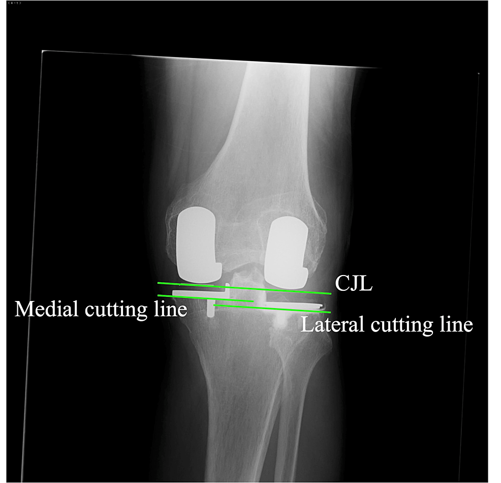
The spoon and the cutting block are fixed using the G-clamp, and the cutting block is fixed using a headless pin (Figure 5, Panel A). After the bone cut and adjustment of the flexion-extension gap are completed on the predominantly affected condyle, the cutting block is removed from the headless pin and changed to that on the opposite side using the same pin along with the extramedullary guide (Figure 5, Panel B) and the ankle yoke with retained extension, side-slide length, and posterior slope to maintain the cutting level as well as sagittal and coronal inclinations. The lesser affected compartment procedure is then performed. When the horizontal cuts are made, insertion of a K-wire at the tip of the tibial spine is recommended to prevent a horizontal overcut (Figure 5, Panel C). After the implantation, the CJL and cutting lines are virtually parallel (Figure 5, Panel D).

Bone-cutting-procedure-for-a-medial-dominant-osteoarthritis

Figure
5:
Bone-cutting procedure for a medial dominant osteoarthritis

(A) After the inclination and level of the cutting block are decided, a headless pin (arrowhead) is inserted into the tibia through the most lateral pinhole of the cutting block. The medial procedure is then performed. (B) The medial cutting block is changed to the lateral cutting block using the same pin connected to the yoke, and the same extent, slide, and posterior slope are maintained. (C) When performing the horizontal cut, a 2 mm K-wire is inserted to prevent a horizontal overcut. (D) The lateral procedure is then performed to set the medial and lateral cutting lines to be parallel.

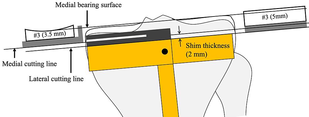
When the lateral tibial cuts are made, the shim is removed so that the cutting level is set to 2 mm lower than the medial cutting level. Both bearings are numbered, but the exact thicknesses of the bearings are 0.5 mm and 2.0 mm thicker than the labeled number. Complete leveling of both plateaus is therefore impossible when the level of the cutting block is constant (Figure 6). Eventually, the lateral CJL is inevitably 0.5 mm higher than the medial CJL, although this can be ignored.

Medial-and-lateral-tibial-cutting-level-in-the-kinematic-alignment-Oxford-unicompartmental-knee-arthroplasty

Figure
6:
Medial and lateral tibial cutting level in the kinematic alignment Oxford unicompartmental knee arthroplasty

The actual thicknesses are 0.5 mm and 2 mm thicker than the labeled number. Once the shim is removed, the lateral cutting level is 2.0 mm lower than that of the medial one. Eventually, the lateral bearing surface is 0.5 mm higher than the medial one when the same number bearing is used with the same cutting block level.

Postoperative radiographical evaluation

True anteroposterior radiography aligned to the tibial component surface is used for postoperative evaluation. Ideally, the postoperative CJL, which is the line tangential to both medial and lateral femoral components, is parallel with the medial and lateral cutting surface, and the medial cutting surface is 2 mm higher than the lateral cutting surface. In straight-type knees on the preoperative radiography, the CJL is expected to be parallel with the preoperative CJL (Figure 7).

Postoperative-radiography

Figure
7:
Postoperative radiography

The coronal joint line (CJL) is parallel to the cutting lines. Note that the lateral cutting line is lower than the medial cutting line.

Discussion

This is the first report to document KA-BiUKA by OPK in detail. Robotic-assisted BiUKA using a fixed-bearing component was recently reported, and constitutional whole leg alignment and joint line obliquity were shown to be restored [19]. Regarding OPK, it was used for BiUKA in the initial stage of the OPK [15]. Pandit et al. reported the staged BiUKA – adding a lateral UKA after medial UKA due to lateral compartmental osteoarthritis and showed satisfactory results [16]. More recently, a gait analysis showed that the subjects with BiUKA using OPK had similar gait characteristics to the normal subject compared to TKA subjects [20]. BiUKA has been reported to have mechanical advantages. A compression force on one component would cause a lift-off of the other component in the one-piece TKA component, but it never occurs in the two-piece tibial components in BiUKA [15,21]. The bone-cutting line was not shown in the previous studies; however, it is thought to play an important role in load transmission. A slight varus implantation of the tibial component was reported in previous biomechanical studies to reduce stress concentration in the medial tibial cortex, but a valgus placement increases it [22]. Although avoidance of valgus placement is important, the placement can be valgus against the proximal tibia in knees with tibia vara, which is especially prevalent in Asian patients [23,24]. Component placement parallel to the CJL might enable a proportioned load transmission across the joint.

In our technique, the dominantly affected compartment is operated upon prior to the lesser affected compartment. In this sequence, the operated condyle is always normal or nearly normal. By contrast, the procedure of the lesser affected condyle can be influenced by the disease of the opposite condyle, such as contracted or relaxed soft tissue and cartilage as well as bone loss. Our technique is a tibia-first sequence, in contrast with most KA-TKA techniques, in which the femur-first technique is used [25,26]. However, this is a standard technique in OPK and has been used for more than 40 years [27]. The tibia-first approach and incremental gap adjustment using the milling system can facilitate easy and precise adjustment of flexion and extension gaps. We believe the dominantly affected condyle-first and the tibial-first sequence might be ideal for facilitating the KA-BiOUKA.

There are some limitations in our report and technique. First, it was necessary to use custom-made devices (accessory spoons and side-slidable ankle yoke). Although a similar operation can be performed without the custom-made devices, where the cutting levels are decided individually using the standard spoon, the cutting plane is not parallel to the CJL. The CJL could be made parallel to the original CJL, but the kinematics and load distribution might be affected. Second, the lateral component is set in varus in the technique. This alignment is equivalent to a valgus placement of the medial UKA, which has reportedly increased the mechanical stress on the tibial cortex. Therefore, it can increase the risk of failure. Varus placement has not been reported to increase the risk of failure in lateral UKA. Third, there was no evaluation of clinical outcomes, in particular its superiority over TKA. A larger number of cases and long-term studies are needed to prove the benefits of the KA-BiUKA. Lastly, we used medial and lateral capsulotomy and disturbance of blood supply for the patella, followed by the avascular necrosis of the patella, and the anterior knee pain is a concern. The medial parapatellar approach for femoral and tibial bone cuts like TKA along with small lateral capsulotomy for lateral gap evaluation using the feeler gauge might be helpful. However, it might require additional instruments.

BiUKA is a great technically demanding operation; therefore, the establishment of the procedure is necessary for a fair evaluation of its effect. Our technique is considered to be easy and reproducible, so it can be implemented widely.

Conclusions

The details of an operative technique of BiUKA using OPK are presented as an alternative procedure for osteoarthritic knees with a functioning ACL and cartilage interaction on the lateral compartments. The technique can replicate the pre-arthritic joint line and maintain both cruciate ligaments, and a cylindrical axis is completely constructed. Moreover, the bone-cutting surface can be set in parallel to the joint line, which might be beneficial to load transmutation.


###
Originally Appeared Here

Filed Under: joint replacement, ORTHO NEWS

Lose Weight, Slow Knee OA Progression? New Study Suggests Yes

by

Declines in body mass index (BMI) were linked with slower worsening of knee osteoarthritis (OA), according to data from three large longitudinal cohort studies.

With more than 6,000 knees evaluated for OA progression and 4 to 5 years of follow-up, “each 1-unit decrease in BMI was associated with a 4.76% reduction … in the odds of the incidence and progression of the overall structural defects of knee osteoarthritis” as assessed under the Kellgren-Lawrence grading system, reported Zubeyir Salis, BEng, of the University of New South Wales in Kensington, Australia, and colleagues in Arthritis & Rheumatology.

While such a relationship seems obvious, it has evaded firm proof, the researchers noted. Earlier studies had tied body weight to risk of OA development and the likelihood of progression and end-stage outcomes such as total joint replacement. But whether losing or gaining weight alters the trajectory over time is another matter.

Salis and colleagues identified just one previous attempt to address the question directly: patients already showing joint degeneration who lost around 10 kg (22 lb) on average in a randomized diet-and-exercise trial showed no less progression than a control group with little weight loss, but follow-up only lasted 18 months. (A number of other studies had lumped patients without structural damage at baseline together with those with established degeneration, and they had mixed results.)

For the current analyses, Salis and colleagues combined data from three independent cohort studies from the U.S. and the Netherlands: the Osteoarthritis Initiative (OAI), which was also the basis for some of the above-mentioned studies; the Multicenter Osteoarthritis Study (MOST); and the Cohort Hip and Cohort Knee (CHECK) study. These data covered 9,683 knees (5,774 individual patients) for assessing the incidence of structural knee degeneration (i.e., no evidence of joint damage at baseline) and 6,074 knees (3,988 individuals) for progression of established degeneration.

Structural joint parameters were measured with x-rays at baseline and at follow-up, which was 4 years in the OAI and 5 years in the other two cohorts. Changes in these parameters (medial and lateral joint space narrowing and femoral and tibial surface osteophytes) were correlated against changes in BMI.

For patients included in the evaluation of new-onset structural damage, mean age at baseline was 60; about 40% were men and 88% were white. BMI at baseline averaged 28.2, with 33% of participants classified as obese (BMI ≥30). A total of 1,101 participants in this analysis saw BMI declines of at least 1 unit during follow-up, whereas 1,611 had increases of 1 or more units.

Patients’ baseline characteristics in the study of progression were similar. Baseline BMI was a bit higher on average (30.4), and 48% were obese. BMI declines of 1 or more units were seen in 798 participants, while 1,008 had increases.

Salis and colleagues found a significant association between BMI changes and the risk of developing structural damage, with an odds ratio of 1.05 (95% CI 1.02-1.09) for each 1-unit increment in BMI. This value was nearly identical to that seen for risk of progression in patients with damage at baseline (OR 1.05, 95% CI 1.01-1.09). In both analyses, joint space narrowing in the medial area largely drove the overall findings, with odds ratios in both cases of 1.08 per 1-unit BMI increment.

However, the results suggested that weight loss was far from the only factor at play in governing joint-damage incidence and progression. The researchers estimated population-attributable fractions of the total risk accounted for by weight loss at 13% for incidence and 10% for progression.

In addition, the investigators stopped short of asserting that the findings prove that weight loss slows OA disease progression, saying they “showed evidence of association, not causality.” But the researchers did argue that “people with overweight or obesity — and potentially also those of normal weight — may benefit from a decrease in BMI to prevent, delay or slow the structural defects in knee osteoarthritis.”

  • John Gever was Managing Editor from 2014 to 2021; he is now a regular contributor.

Disclosures

This study was supported by the Australian government; the individual cohort studies each had their own funding sources, none of which were commercial entities.

Salis and one co-author were co-owners of a company dedicated to education around weight management. Two co-authors also reported relationships with multiple pharmaceutical companies.

###
Originally Appeared Here

Filed Under: joint replacement, ORTHO NEWS

Consultant offers reduced waiting times and better outcomes for hip and knee surgery

by

With NHS waiting times for hip and knee replacements in the North East and North Yorkshire currently up to two years, Mr Andrew Port, a highly experienced consultant orthopaedic surgeon, is offering the people of these regions quicker access for their joint replacement surgery.

Mr Port offers his patients individualised care pathways for hip and knee replacements, including partial knee replacements, complex and revision (redo) hip and knee replacements, for which he is a regional specialist.

As a pioneer for robotic joint replacement surgery in the UK, his patients are now able to take advantage of the quicker recovery times, reduced pain and improved function, following robotic joint replacement surgery.


His current waiting time for a joint replacement privately is four weeks, and for and NHS patient is around six months.

Mr Port has more than 30 years’ experience in orthopaedic surgery, during which time he has led and guided local, regional and national directives in improving the quality of orthopaedic surgery. He has been based at the BMI Woodlands hospital in Darlington for 22 years.

The Northern Echo:

He performs more than 500 hip and knee replacements a year, including 50 revision surgeries and 40 partial knee replacements. The “Getting It Right First Time” (GIRFT) Department of Health directive and the British Orthopaedic Association advocate that surgeons should perform a minimum of 15 of each of these procedures per year to maintain standards. Mr Port has one of the highest patient satisfaction outcomes and lowest complication rates nationally.

The Northern Echo:

Being one of the first surgeons in the UK to introduce robotic hip and knee replacement surgery, Mr Port has performed more than 300 robotic-assisted hip and knee replacements using the Stryker Mako Robotics system.

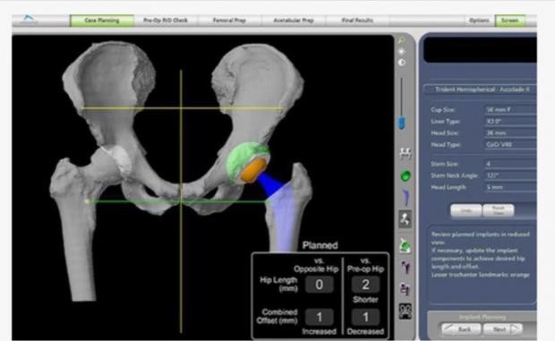
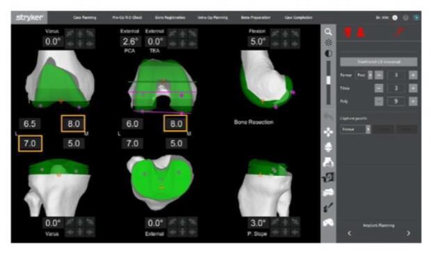
From his experience he explains: “The technology combines 3D planning with accurate intra-operative reconstruction of the hip or knee replacement. Following robotic surgery, patients are reporting less pain, quicker recovery times and greater satisfaction rates”.

The Northern Echo:

The BMI Woodlands Hospital, in Darlington, is the only hospital in the northern region that offers access to augmented surgical assistance with the Stryker Mako Robotics system.

Mr Port is currently chairman of the Surgical Collaborative at South Tees NHS Trust, incorporating The James Cook University Hospital and the Friarage Hospital. He leads on the strategy to standardise the delivery and quality of orthopaedic surgery over the Tees Valley and North Yorkshire.

Further information can be found at: www.circlehealthgroup.co.uk/consultants/andrew-port or, to book an appointment, contact 01325 341784, or Mr Port’s practice manager on 07855 364475.


###
Originally Appeared Here

Filed Under: joint replacement, ORTHO NEWS

‘Covid recovered patients prone to orthopaedic complications’

by

Published: Published Date – 08:56 PM, Sun – 11 September 22

‘Covid recovered patients prone to orthopaedic complications’
(Representational Image) Covid-19 recovered patients are prone to orthopaedic complications like osteonecrosis of the hip, which damages hip joint, senior orthopaedic surgeons said at AAS Summit-2022.

Hyderabad: Covid-19 recovered patients are prone to orthopaedic complications like osteonecrosis of the hip, which damages hip joint and could also cause severe arthritis, senior orthopaedic surgeons at a Arthroplasty Arthroscopy Summit-2022 (AAS), organised by Apollo Hospitals, Secunderabad, and Apollo Institute of Medical Sciences and Research (AIMSR), on Sunday, said.

Cases of osteonecrosis are being reported among individuals aged between 20 years and 30 years. “Hip replacement is a gold-standard for treating necrosis. However, since a majority of the patients are from a young age group, hip replacement is usually not recommended. There are modern techniques that provide safe alternatives to such patients,” senior joint replacement surgeons from Apollo Hospitals, Dr Aachi Mithin and Dr N Somashekhar Reddy, said.

In the training program, the faculty highlighted new therapeutics including pain portal injections, which involves injecting of safe local medications in small quantities at various spots to produce long lasting relief to patients. Such injections are giving good midterm to long term relief from pain, Dr L Prakash, Director and Chief of Orthopaedics, Institute for Special Orthopaedics, Palakkad, Kerala, said.

###
Originally Appeared Here

Filed Under: joint replacement, ORTHO NEWS

  • « Go to Previous Page
  • Go to page 1
  • Go to page 2
  • Go to page 3
  • Go to Next Page »

Primary Sidebar

ORTHO NEWS

Spinal anesthesia tied to increased opioid use after hip surgery

Spinal anesthesia tied to increased opioid use after hip surgery

Useful Health Tips for People With Chronic Hip Issues

Useful Health Tips for People With Chronic Hip Issues

A robotic assistant for joint replacement surgery

A robotic assistant for joint replacement surgery

Prior Diagnosis of COVID Has No Increased Complications in Total Joint Arthroplasty

Prior Diagnosis of COVID Has No Increased Complications in Total Joint Arthroplasty

Putting Your Best Foot Forward for Joint Surgery Success | Health

Putting Your Best Foot Forward for Joint Surgery Success

Use of Antibiotic Lavage in Total Knee Replacement to Prevent Postoperative Infection

Use of Antibiotic Lavage in Total Knee Replacement to Prevent Postoperative Infection

Tips to care for an elderly after hip replacement surgery

Tips to care for an elderly person after hip replacement surgery

Exactech Total Knee Failure Resulted in Revision Surgery, Lawsuit Alleges

Exactech Total Knee Failure Resulted in Revision Surgery, Lawsuit Alleges

AAOS Updates Clinical Practice Guideline for Surgical Management of Osteoarthritis of the Knee

AAOS Updates Clinical Practice Guideline for Surgical Management of Osteoarthritis of the Knee

Robotics Procedure Helping People Recover From Joint Replacement Surgery

Robotics Procedure Helping People Recover From Joint Replacement Surgery

Copyright 2014 All Rights Reserved · DISCLAIMER: Nothing here constitutes legal, medical, or other advice; all content relates to an individual perspective only. A professional relationship with a physician, or with a lawyer is built over time, with mutual investment, trust, and respect. This site is not a substitute for that.
~ THIS DOMAIN IS FOR SALE ~

Privacy Policy