Arthritis of the knee is a common underlying cause for knee replacement surgery. Here’s what you should know.
How is worn cartilage diagnosed?
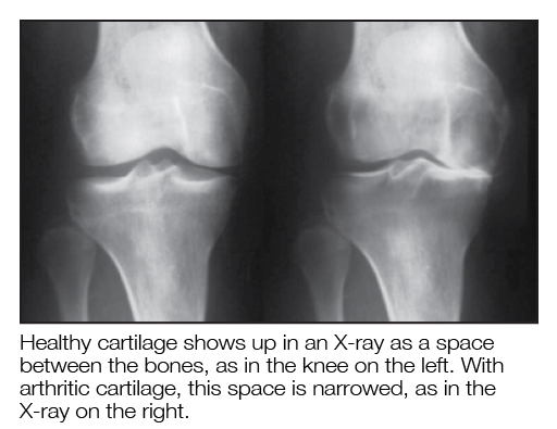
Symptoms such as pain, swelling, stiffness, lack of movement, and grinding in the knee can suggest that knee cartilage is no longer smooth. X-rays are a common way of diagnosing diseased cartilage in the knee. The X-rays show the bones around the joint. The cartilage layer cannot be seen on X-rays, and shows up as a space between the femur and tibia bones.

In a healthy knee, this space is about a quarter of an inch thick. When joint space taken up by cartilage is destroyed by arthritis, X-rays will show joint-space narrowing. With significant cartilage loss, the bones may touch each other; doctors call this finding “bone-on-bone” on the X-rays.
Over time, cysts and bone spurs may form around the knee. Left untreated, the leg can get so deformed that it appears to be either bow-legged or knock-kneed.
Are there other ways to see the extent of damage to knee cartilage?
MRI scans are a special X-ray study that can diagnose diseased cartilage somewhat more accurately and at an earlier point than plain X-rays. Another method includes actually looking inside the knee during a procedure called “knee arthroscopy.” This involves the surgeon placing a small camera in the knee and inspecting the cartilage.